| Term | Definition |
|---|---|
| Biology | The study of life and living organisms. |
| Botany | The study of plants. |
| Zoology | The study of animals. |
| Chemistry | The study of the composition and properties of substances. |
| Physics | The study of matter, energy and their interactions. |
| Microbiology | The study of microorganisms (organisms too small to be seen with the naked eye). |
| Bacteriology | The study of bacteria. |
| Virology | The study of viruses. |
| Mycology | The study of fungi. |
| Cytology | The study of cells. |
| Histology | The study of tissues (the microscopic structure of tissues). |
| Genetics | The study of heredity and genes. |
| Ecology | The study of the relationships between organisms and their environment. |
| Anatomy | The study of the internal structure of the body. |
| Physiology | The study of the normal functions of the body and its parts. |
| Pathology | The study of diseases (their nature, causes and effects). |
| Pharmacology | The study of drugs and their actions on the body. |
| Entomology | The study of insects. |
| Ichthyology | The study of fish. |
| Ornithology | The study of birds. |
| Herpetology | The study of reptiles and amphibians. |
| Mammalogy | The study of mammals. |
| Cardiology | The study of the heart and its diseases. |
| Neurology | The study of the nervous system (brain, spinal cord and nerves). |
| Nephrology | The study of the kidneys. |
| Hepatology | The study of the liver. |
| Dermatology | The study of the skin. |
| Ophthalmology | The study of the eyes. |
| Haematology | The study of blood. |
| Oncology | The study of tumours and cancer. |
| Immunology | The study of the immune system. |
| Epidemiology | The study of the distribution and control of diseases in populations. |
| Astronomy | The study of celestial (heavenly) bodies. |
| Geology | The study of the earth, its structure and rocks. |
| Geography | The study of the earth's surface, its features and inhabitants. |
| Meteorology | The study of the weather and the atmosphere. |
| Seismology | The study of earthquakes. |
| Oceanography | The study of the oceans. |
| Psychology | The study of the mind and behaviour. |
| Sociology | The study of human society. |
| Biochemistry | The study of the chemical processes within living organisms. |
| Taxonomy | The study of the classification of organisms. |
| Term | Definition |
|---|---|
| Parasitology | The study of parasites and their hosts. |
| Helminthology | The study of parasitic worms (helminths). |
| Protozoology | The study of protozoa (single-celled animal-like organisms). |
| Acarology | The study of mites and ticks. |
| Ethology | The study of animal behaviour. |
| Phycology | The study of algae (also called Algology). |
| Ophiology | The study of snakes. |
| Arachnology | The study of spiders and other arachnids. |
| Cetology | The study of whales, dolphins and other cetaceans. |
| Embryology | The study of the embryo and its development. |
| Palaeontology | The study of fossils and ancient life. |
| Toxicology | The study of poisons and their effects on the body. |
| Gynaecology | The study of the female reproductive system and its diseases. |
| Obstetrics | The branch of medicine dealing with pregnancy, childbirth and midwifery. |
| Paediatrics | The branch of medicine dealing with the health and diseases of children. |
| Endocrinology | The study of the endocrine glands and their hormones. |
| Gastroenterology | The study of the stomach and intestines (the digestive system). |
| Osteology | The study of bones. |
| Myology | The study of muscles. |
| Otology | The study of the ear. |
| Odontology | The study of teeth. |
| Urology | The study of the urinary system and its diseases. |
| Pulmonology | The study of the lungs and the respiratory system. |
| Radiology | The study of X-rays and medical imaging. |
| Psychiatry | The study and treatment of mental disorders. |
| Gerontology | The study of ageing and old age. |
| Agronomy | The study of soil management and crop production. |
| Pedology | The study of soils (their formation and classification). |
| Hydrology | The study of water on the earth (its distribution and movement). |
| Climatology | The study of climate (long-term weather patterns). |
| Limnology | The study of inland fresh waters (lakes, rivers and ponds). |
| Vulcanology | The study of volcanoes (also spelled Volcanology). |
| Archaeology | The study of human history through material remains. |
| Anthropology | The study of human beings — their origin, development and cultures. |
| Demography | The study of human populations and their statistics. |
| Cartography | The science of making maps. |
| Numismatics | The study of coins and currency. |
| Etymology | The study of the origin and history of words. |
| Linguistics | The study of language. |
| Pomology | The study of fruits and their cultivation. |
| Horticulture | The cultivation of garden plants (fruits, vegetables, flowers and ornamental plants). |
| Agriculture | The science and practice of farming — cultivation of land and rearing of livestock. |
| Sericulture | The rearing of silkworms for the production of silk. |
| Apiculture | The rearing of honey bees (bee-keeping). |
| Pisciculture | The rearing of fish (fish farming). |
| Anaesthesiology | The study of anaesthesia and anaesthetic agents. |
| Rheumatology | The study of rheumatic diseases (of joints, muscles and connective tissue). |
| Serology | The study of blood serum, especially its antibody (immune) reactions. |
| Primatology | The study of primates (monkeys, apes and man). |
| Term | Definition |
|---|---|
| Theriogenology | The study of animal reproduction (veterinary obstetrics and gynaecology). |
| Epizootiology | The study of disease patterns and outbreaks in animal populations. |
| Andrology | The study of the male reproductive system and male infertility. |
| Nematology | The study of nematodes (roundworms). |
| Malacology | The study of molluscs (snails, slugs, oysters etc.). |
| Conchology | The study of shells (of molluscs). |
| Lepidopterology | The study of butterflies and moths. |
| Coleopterology | The study of beetles and weevils. |
| Apiology | The study of honey bees (genus Apis). The study of bees in general is melittology. |
| Dendrology | The study of trees. |
| Bryology | The study of mosses and liverworts (bryophytes). |
| Lichenology | The study of lichens. |
| Agrostology | The study of grasses. |
| Petrology | The study of rocks (their origin, composition and structure). |
| Mineralogy | The study of minerals. |
| Geomorphology | The study of landforms and the earth's surface features. |
| Glaciology | The study of ice and glaciers. |
| Speleology | The study of caves. |
| Selenology | The study of the moon. |
| Cosmology | The study of the origin and structure of the universe. |
| Astrophysics | The study of the physical nature of celestial bodies. |
| Chronology | The science of arranging events in their order of time. |
| Genealogy | The study of family descent and ancestry. |
| Heraldry | The study of coats of arms. |
| Philately | The study and collection of postage stamps. |
| Epigraphy | The study of inscriptions (on stone, metal etc.). |
| Lexicology | The study of words and their meanings (vocabulary). |
| Phonology | The study of the sound system of a language (sounds of speech). |
| Semantics | The study of meaning in language. |
| Theology | The study of religion and the nature of God. |
| Mythology | The study of myths and traditional tales. |
| Criminology | The study of crime and criminals. |
| Penology | The study of the punishment of crime and the management of prisons. |
| Rhinology | The study of the nose. |
| Laryngology | The study of the larynx (voice box) and throat. |
| Nosology | The study of the classification of diseases. |
| Aetiology | The study of the causes of diseases (also spelled Etiology). |
| Neonatology | The branch of medicine dealing with newborn infants. |
| Kinesiology | The study of body movement and posture. |
| Arthrology | The study of joints. |
| Angiology | The study of the blood vessels and the lymphatic system. |
| Viticulture | The cultivation of grapevines (grapes). |
| Floriculture | The cultivation of flowers (flower farming). |
| Olericulture | The cultivation of vegetables. |
| Silviculture | The cultivation and management of forest trees. |
| Aquaculture | The farming of aquatic organisms (fish, prawns and aquatic plants). |
| Vermiculture | The rearing of earthworms (for vermicompost). |
| Aviculture | The rearing and breeding of birds. |
| Zymology | The study of fermentation. |
| Oenology | The study of wines and wine-making. |
| Gemmology | The study of gems and precious stones. |
| Metallurgy | The study of metals — their extraction, refining and use. |
| Acoustics | The study (science) of sound. |
| Optics | The study of light and its behaviour. |
| Thermodynamics | The study of heat and its relation to energy and work. |
| Term | Definition |
|---|---|
| Onychology | The study of nails (and claws in animals). |
| Hippology | The study of horses. |
| Cynology | The study of dogs. |
| Felinology | The study of cats. |
| Chiropterology | The study of bats. |
| Myrmecology | The study of ants. |
| Pteridology | The study of ferns. |
| Palynology | The study of pollen and spores. |
| Pharmacognosy | The study of medicinal drugs obtained from plants and other natural sources. |
| Teratology | The study of congenital abnormalities and abnormal development (birth defects). |
| Symptomatology | The study of the symptoms of diseases. |
| Phlebology | The study of veins. |
| Trichology | The study of hair and the scalp. |
| Somnology | The study of sleep. |
| Thanatology | The study of death and dying. |
| Vexillology | The study of flags. |
| Campanology | The study of bells and bell-ringing. |
| Graphology | The study of handwriting. |
| Oneirology | The study of dreams. |
| Areology | The study of the planet Mars. |
| Hippiatrics | The study of the diseases of horses (veterinary medicine of horses). |
| Arboriculture | The cultivation and management of individual trees and shrubs. |
| Mariculture | The farming of marine organisms (sea fish, oysters etc.) in the sea. |
| Bromatology | The study of food. |
| Edaphology | The study of soil in relation to plant growth. |
| Cryobiology | The study of the effects of low temperatures on living organisms. |
| Aerobiology | The study of airborne organisms and particles (pollen, spores and microbes in the air). |
| Exobiology | The study of life beyond the earth (also called Astrobiology). |
| Ethnology | The study of human races and cultures (the comparative study of peoples). |
| Dendrochronology | The study of the age of trees from their annual growth rings. |
| Karyology | The study of cell nuclei. |
| Ichnology | The study of fossil footprints and other traces of animals. |
| Hyetology | The science of rainfall. |
| Nephology | The study of clouds. |
| Oology | The study of eggs (especially birds' eggs). |
| Caliology | The study of birds' nests. |
| Desmology | The study of ligaments. |
| Splanchnology | The study of the internal organs (viscera). |
| Enzymology | The study of enzymes. |
| Histopathology | The study of diseased tissues (tissue changes caused by disease). |
| Phytopathology | The study of plant diseases. |
| Anemology | The study of winds. |
| Heliology | The science of the sun. |
| Geodesy | The science of measuring the earth's shape and size. |
| Topography | The study and description of the surface features of a place. |
| Horology | The science of measuring time (and of making clocks and watches). |
| Dactylography | The study of fingerprints (for identification). |
| Iamatology | The study of remedies (medicines and cures). |
| Particulars | Cattle | Buffalo | Sheep | Goat | Horse |
|---|---|---|---|---|---|
| Rectal Temperature (°F) | 101.5 | 101.0 | 103.0 | 103.0 | 100.5 |
| Normal Pulse Rate/min | 50-70 | 48-65 | 70-80 | 70-80 | 30-40 |
| Normal Resp. Rate/min | 20-30 | 20-28 | 20-30 | 20-30 | 10-16 |
| Age at First Service (mo) | 15-18 | 26-30 | 8-12 | 8-12 | ~36 |
| Gestation Period (days) | 283 | 304 | 150 | 150 | 340 |
| Particulars | Dog | Cat | Camel | Fowl |
|---|---|---|---|---|
| Rectal Temperature (°F) | 101-102 | 101.0 | 99.5 | 107.0 |
| Normal Pulse Rate/min | 70-130 | 110-130 | 30-50 | 250–300 |
| Normal Resp. Rate/min | 15-30 | 20-30 | 10-12 | 15-30 |
| Age at First Service (mo) | 6-12 | 6-15 | -- | -- |
| Gestation Period (days) | 63 | 63–65 | 370 | 21 (hatch) |
| Common Name | Scientific Name |
|---|---|
| Alpaca | Lama pacos (syn. Vicugna pacos) |
| Ass | Equus asinus |
| Buffalo | Bubalus bubalis |
| Camel | Camelus dromedarianus (Single hump); Camelus bactrianus (Double hump) |
| Cat | Felis catus |
| Cattle | Bos indicus / Bos taurus |
| Dog | Canis familiaris |
| Goat | Capra hircus |
| Guanaco | Lama guanicoe |
| Horse | Equus caballus |
| Llama | Lama glama |
| Mithun | Bos frontalis |
| Pig | Sus scrofa |
| Rabbit | Oryctolagus cuniculus |
| Reindeer | Rangifer tarandus |
| Sheep | Ovis aries |
| Vicuna | Vicugna vicugna |
| Yak | Bos grunniens |
| Common Name | Scientific Name |
|---|---|
| Chicken | Gallus domesticus |
| Duck | Anas platyrhynchos |
| Geese | Anser anser |
| Golden eagle | Aquila chrysaetos |
| Guinea fowl | Numida meleagris |
| Muscovy | Cairina moschata |
| Ostrich | Struthio camelus |
| Pea fowl (peacock) | Pavo cristatus |
| Pigeon | Columba livia |
| Pheasants | Phasianus colchicus |
| Quail | Coturnix spp. |
| Eurasian eagle-owl | Bubo bubo |
| Barn owl | Tyto alba |
| Swan | Cygnus spp. |
| Turkey | Meleagris gallopavo |
Vertical transmission means the infection passes from the parent — almost always the dam — to her offspring: across the placenta before birth, through the egg in poultry, at birth, or in the first colostrum and milk. The disease jumps a whole generation without ever spreading in the shed, so a single infected breeder can seed an entire line of animals.
| Route | How it happens | Typical examples |
|---|---|---|
| Transplacental (in utero) | The agent crosses the placenta and infects the fetus during pregnancy | BVD, Neospora, toxoplasmosis, border disease |
| Transovarial (true egg transmission) | The ovary or oviduct is infected, so the agent is already inside the egg before it is laid | Pullorum disease, Mycoplasma, avian leukosis |
| Egg-shell contamination (false vertical) | Bacteria on the shell (from feces) penetrate into the egg after laying | Salmonella, E. coli (omphalitis) |
| Through colostrum and milk | The agent is shed in colostrum/milk and infects the newborn at its first suckling | CAEV, maedi-visna, BLV |
| Perinatal (at birth) | The offspring is infected passing through the birth canal or from genital discharges at birth | Canine herpesvirus, enzootic abortion of ewes |
| Disease | Agent | Vertical route | Key point |
|---|---|---|---|
| Bovine viral diarrhea (BVD) | Pestivirus | Transplacental | Infection early in pregnancy produces a PERSISTENTLY INFECTED (PI) calf — the main reservoir of the virus |
| Neosporosis | Neospora caninum (protozoon) | Transplacental | Dog is the definitive host; a leading infectious cause of abortion in cattle, passed cow-to-calf very efficiently |
| Toxoplasmosis | Toxoplasma gondii | Transplacental | Cat is the definitive host; a major cause of abortion in sheep and goats |
| Border disease | Pestivirus | Transplacental | PI lambs are born weak with a tremor — the “hairy shaker” lamb |
| Bluetongue | Orbivirus | Transplacental (some strains) | Main spread is horizontal by Culicoides midges; crossing of the placenta was shown especially with BTV-8 |
| Schmallenberg disease | Orthobunyavirus (family Peribunyaviridae) | Transplacental | A midge infects the dam; the damage — deformed lambs and calves — is done inside the fetus |
| Akabane disease | Orthobunyavirus (Simbu group) | Transplacental | A midge infects the dam; the fetus is born with arthrogryposis (fixed joints) and hydranencephaly (cerebral hemispheres missing) |
| Brucellosis | Brucella abortus | In utero / at birth | Calves of infected cows may carry a latent infection that only shows at their first calving |
| Enzootic abortion of ewes | Chlamydia abortus | Perinatal (at lambing) | Ewes infected at one lambing abort at the next; lambs can be born infected |
| PRRS | Arterivirus | Transplacental (late gestation) | Crosses the placenta late in pregnancy — abortions, stillborn and weak piglets |
| Porcine parvovirus (SMEDI) | Parvovirus | Transplacental | Kills embryos and fetuses at successive stages — the litter shows mummified fetuses of different sizes |
| CAEV and maedi-visna | Lentiviruses | Colostrum and milk | Kids and lambs are infected at the first suckling; control is snatch-rearing on pasteurised colostrum |
| Bovine leukosis (BLV) | Retrovirus | In utero (minor) and colostrum/milk | Most spread is iatrogenic/horizontal, but a minority of calves are already born infected |
| Johne’s disease | Mycobacterium avium subsp. paratuberculosis | In utero (advanced cases) and milk/colostrum | Infection picked up at birth decides the wasting disease seen years later |
| Feline leukaemia (FeLV) | Retrovirus | Transplacental and milk | Kittens of infected queens are born infected or pick it up at suckling |
| Feline panleukopenia | Parvovirus | Transplacental | In-utero infection destroys the developing cerebellum — kittens are born wobbly and uncoordinated (cerebellar hypoplasia) |
| Canine herpesvirus infection | Herpesvirus | Perinatal (birth canal) | Cause of “fading puppy” deaths in the first three weeks of life |
| Disease | Agent | Key point |
|---|---|---|
| Pullorum disease | Salmonella Pullorum | The CLASSIC transovarial disease — the infected hen’s ovary seeds the egg; white diarrhoea in chicks |
| Fowl typhoid | Salmonella Gallinarum | Egg-borne like pullorum, but mainly a disease of adult birds |
| Mycoplasmosis (CRD) | Mycoplasma gallisepticum / M. synoviae | The MOST COMMON egg-transmitted disease of poultry; it also spreads horizontally |
| Avian leukosis | Avian leukosis virus (retrovirus) | Congenital transmission through the egg is the main route to the tumours; horizontal spread is slow |
| Reticuloendotheliosis | Reticuloendotheliosis virus (retrovirus) | Passed vertically through the egg/embryo; an immunosuppressive virus causing runting and tumours |
| Avian encephalomyelitis | Picornavirus | The hen passes the virus through her eggs for a few weeks; chicks show “epidemic tremor” |
| Egg drop syndrome (EDS-76) | Adenovirus | Travels vertically in the egg, stays latent, and switches on when the hen comes into lay |
| Inclusion body hepatitis (IBH) | Fowl adenoviruses (FAdV-2, 8, 11) | Egg transmission is documented from infected breeder flocks (also horizontal); sudden deaths in young broilers with an inflamed liver |
| Hydropericardium syndrome (HPS) | Fowl adenovirus 4 (FAdV-4) | Egg transmission is documented (also horizontal); straw-coloured fluid fills the pericardial sac — mortality can reach 20–80% in young broilers |
| Chicken infectious anemia | Gyrovirus — chicken anemia virus (family Anelloviridae) | Seronegative hens pass the virus through the egg; the chicks hatch with NO maternal antibody and show disease at about 2–3 weeks of age |
| Avian reovirus infections | Reovirus | Egg transmission is reported; horizontal spread also occurs |
| Paratyphoid salmonellosis | Salmonella Enteritidis and others | Contaminate the egg contents and shell — the food-safety route of Salmonella to man |
| Colibacillosis (omphalitis) | E. coli | Mostly shell contamination after laying (false vertical) — yolk-sac infection of the newly hatched chick |
Horizontal transmission is the everyday spread of infection between animals of the same population — by contact, through the air, in feed and water, on fomites, or by vectors. It is the route that turns one sick animal into a shed-wide outbreak, and it is the route that biosecurity is designed to stop.
| Mode | How it happens | Examples |
|---|---|---|
| Direct contact | Body contact, licking, fighting, nosing between animals | Ringworm, mange, sheep pox |
| Droplet / aerosol | Coughing and breathing — droplets over a short distance, fine airborne particles over a long one | FMD, IBR, Newcastle disease |
| Feed and water (oral) | Contaminated feed, water, litter or soil is swallowed | Anthrax spores from soil, coccidiosis from litter |
| Fomites | The agent rides on boots, equipment, vehicles, hands or milking clusters | IBD (Gumboro), FMD, contagious mastitis |
| Vectors | Biting flies, ticks or midges carry the agent — mechanically or biologically | Bluetongue (midge), babesiosis (tick), LSD (flies) |
| Venereal | At coitus, or through infected semen and AI | Genital IBR, canine brucellosis, equine coital exanthema |
| Iatrogenic | Man carries it — shared needles, surgical instruments, rectal sleeves | BLV, equine infectious anemia |
| Wounds and skin | The agent enters through bites, wounds or broken skin | Rabies (bite); black quarter in sheep (soil into wounds) — in cattle its spores are swallowed and lie latent in muscle |
| Disease | Agent | Main horizontal mode |
|---|---|---|
| Foot-and-mouth disease | Aphthovirus | Aerosol, direct contact and fomites — among the most contagious diseases of all |
| Haemorrhagic septicaemia | Pasteurella multocida | Droplets and direct contact, typically after stress or rain |
| Lumpy skin disease | Capripoxvirus | Biting flies as mechanical vectors; also direct contact |
| Peste des petits ruminants (PPR) | Morbillivirus | Close contact and respiratory droplets |
| Sheep pox / goat pox | Capripoxvirus | Direct contact, aerosols and fomites |
| Tuberculosis | Mycobacterium bovis | Aerosol — inhaled between cattle in shared air space |
| Strangles | Streptococcus equi | Direct contact, shared water troughs and fomites (horses) |
| Contagious mastitis | Staphylococcus aureus, Streptococcus agalactiae | At milking — hands, cloths and clusters carry it from teat to teat |
| Ringworm (dermatophytosis) | Trichophyton / Microsporum fungi | Direct contact and fomites such as grooming kit and bedding |
| Mange | Sarcoptes and other mites | Direct body contact |
| Rabies | Lyssavirus | Bite of a rabid animal — saliva into the wound |
| Babesiosis and theileriosis | Tick-borne protozoa | Tick bite (biological vector) |
| Equine infectious anemia | Lentivirus | Horse flies (mechanical) and shared needles (iatrogenic) |
| Disease | Agent | Main horizontal mode |
|---|---|---|
| Newcastle disease | Paramyxovirus | Aerosol, direct contact and fomites — very fast shed-wide spread |
| Avian influenza | Orthomyxovirus | Droplets, direct contact, wild waterfowl and fomites |
| Infectious bronchitis | Coronavirus | Aerosol — can sweep through a house within days |
| Infectious laryngotracheitis | Herpesvirus | Aerosol and direct contact |
| Marek’s disease | Herpesvirus | Feather dander and dust in the air — NOT through the egg |
| Infectious bursal disease (Gumboro) | Birnavirus | Fomites, litter, feed and water — the virus survives for months in the shed |
| Fowl cholera | Pasteurella multocida | Direct contact, contaminated water, wild birds and rodents |
| Coccidiosis | Eimeria species | Sporulated oocysts picked up from the litter (oral–environmental) |
| Aspergillosis | Aspergillus fumigatus | Spores inhaled from moldy litter or feed — “brooder pneumonia” |
| Point | Vertical | Horizontal |
|---|---|---|
| Direction | Parent → offspring | Animal → animal (same generation) |
| Timing | Before birth, at birth, in the egg, or at first suckling | Any time after birth or hatching |
| Classic examples | BVD (PI calf), pullorum disease, neosporosis | FMD, Newcastle disease, coccidiosis |
| Control focus | The breeding stock — testing, culling, egg and colostrum hygiene | The population — biosecurity, quarantine, ventilation, vector control, vaccination |
| Name of Disease | Synonym | Causative Agent |
|---|---|---|
| Foot and Mouth Disease | FMD; Apthous fever | Apthovirus, Picornaviridae |
| Rinderpest | Cattle plague; Bovine typhus | Rinderpest virus of family Paramyxoviridae |
| Peste Des Petitis Ruminants | PPR; Pseudorinderpest, Kata, Goat plague | Morbillivirus of family Paramyxoviridae |
| Bovine Viral Diarrhea | BVD; Mucosal disease | Pestivirus of family Flaviviridae |
| Malignant Catarrhal Fever | MCF; Catarrhal fever; Gangrenous coryza | Gamma herpesvirus |
| Blue Tongue | Sore mouth; Epizootic catarrh; Pseudo FMD | Orbivirus of family Sedoreoviridae (formerly Reoviridae) |
| Vesicular Stomatitis | Mouth thrush; Sporadic aptha; Estomatitis | VS virus of family Rhabdoviridae |
| Ephemeral Fever | Three day sickness; Stiff sickness | EF virus of family Rhabdoviridae |
| Infectious Bovine Rhinotracheitis | IBR; Red nose; Necrotic rhinitis | Bovine herpes virus-I |
| Pseudo rabies | Aujeszky's disease; Mad itch | Herpes virus |
| Cow pox | Variola vaccina | Cow pox virus |
| Goat pox | Variola capra | Capri pox virus |
| Sheep pox | Ovine px; Variola ovina | Capri pox virus |
| Swine pox | Variola suilla; Contagious impetigo | Sui pox virus |
| Horse pox | Variola equine; Contagious pustular stomatitis | Equi pox virus |
| Pseudo cow pox | Milker's nodules; Ring sore | Para pox virus |
| Swine Fever | Hog cholera; Peste porcine | Pestivirus of family Flaviviridae |
| Vesicular Exanthema | -- | Calicivirus |
| African Swine Fever | African pig disease; Wart hog disease | Asfivirus of family Asfarviridae |
| Transmissible Gastroenteritis | T.G.E. | Corona virus |
| Japanese Encephalitis | -- | Flavivirus of family Flaviviridae |
| Contagious Ecthyma | Contagious pustular dermatitis; ORF; Scabby mouth | ORF virus of family Poxviridae |
| Maedi | Progressive interstitial pneumonia | Lentivirus of family Retroviridae |
| Pulmonary Adenomatosis | Jaagsiekte | Jaagsiekte sheep retrovirus (JSRV) of family Retroviridae |
| Bovine Leukosis | Bovine lymphosarcoma | Bovine leukemia virus of family Retroviridae |
| Corona virus infection | CCV; Viral gastroenetritis | Corona virus |
| Equine Influenza | Equine distemper; Typhoid fever; Pink eye | Influenza A virus (Orthomyxoviridae) |
| Equine Infectious Anemia | EIA; Swamp fever | EIA virus; Lentivirus of family Retroviridae |
| Respiratory Disease Complex in Dog | Kennel cough | Bordetella bronchiseptica (a bacterium — multifactorial complex) |
| Equine Viral Rhinopneumonia | Rhinopneumonitis; Equine viral abortion | Equine herpes virus 1 and 4 (EHV-1, EHV-4) |
| Scrapie | Rida; Tremblante du mouton | Prion |
| Louping ill | Ovine encephalomyelitis | Flavivirus of family Flaviviridae |
| Bovine Spongiform Encephalopathy | BSE; Mad cow disease | Prion |
| Canine Distemper | Hard pad disease; Hard pad disease | Canine distemper virus of family Paramyxoviridae |
| Infectious Canine Hepatitis | Rubarth's disease; Contagious hepatitis | Canine adenovirus 1 (CAV-1) of family Adenoviridae |
| Rabies | Mad dog; Hydrophobia; Jalatanka; Lyssa | Lyssa virus of family Rhabdoviridae |
| Rotavirus Diarrhea | -- | Genus Rotavirus of family Sedoreoviridae |
| Parvovirus Infection in Dog | -- | Parvovirus of family Parvoviridae |
| Feline Panleukopenia | Feline distemper; Feline parvovirus; Feline ataxia | DNA virus |
| Respiratory Disease Complex in Cat | Cat flu, FVR | Feline herpes virus-I |
| Feline Leukemia | Lymphosarcoma | Retrovirus |
| Name of disease | Synonym | Causative Agent |
|---|---|---|
| Anthrax | Splenic fever; Charbon; Wool sorter's disease | Bacillus anthracis |
| Hemorrhagic Septicemia | Pasteurellosis; Shipping fever; Stockyard disease | Pasteurella multocida |
| Brucellosis | Bang's disease; Infectious abortion; Enzootic abortion | B.abortus, B. melitensis, B. suis, B. ovis, B. canis |
| Tuberculosis | Pearl's disease; Pthisis; Scrofula | Mycobacterium spp. |
| Paratuberculosis | Johne's disease; Chronic bacillary dysentery | Mycobacterium avium subsp. paratuberculosis (MAP; formerly Mycobacterium paratuberculosis) |
| Leptospirosis | Weil's disease; Stuttgart disease; Rice field worker's disease; Canine typhus | Leptospira spp. (L.interrogans, L.pomona, L.canicola, L.hardjo) |
| Listeriosis | Circling disease; Silage disease | Listeria monocytogenes |
| Colibacillosis | Calf scour; White scour; Calf septicemia | Eschericia coli (E.coli) |
| Salmonellosis | Paratyphoid | S. typhimurium, S.dublin |
| Actinomycosis | Lumpy jaw | Actinomyces bovis |
| Actinobacillosis | Wooden tongue; Timber tongue | Actinobacillus lignieresii |
| Dermatophylosis | Mycotic dermatitis; Senekobo disease of cattle, Lumpy wool of sheep | Dermatophilus congolensis |
| Swine Erysipelas | Diamond skin disease | Erysipelothrix rhusiopathiae |
| Compylobacteriosis | Vibriosis | Campylobacter foetus |
| Glanders | Farcy; Malleus; Morve; Pacin; Carn | Burkholderia mallei |
| Strangles | Equine distemper; Infectious adenitis | Streptococcus equi |
| Exudate Epidermitis | Greasy pig disease | Staphylococcus hyicus |
| Impetigo | -- | Staphylococcus aureus |
| Glasser's disease | Infectious polyarthritis | Glaesserella parasuis |
| Ulcerative Lymphangitis | Ulcerative cellulitis; — (CLA is a separate sheep/goat syndrome) | Corynebacterium pseudotuberculosis |
| Foot Rot | Infectious pododermatitis | Fusobacterium necrophorum |
| Infectious Kerato Conjunctivitis | Pink eye; Infectious keratitis | Moraxella bovis |
| Black Quarter | Black leg; Quarter ill; Symptomatic anthrax | Clostridium chauvoei |
| Tetanus | Lock jaw | Clostridium tetani |
| Enterotoxemia | -- | Clostridium perfringens |
| Bacillary Hemoglobinurea | Red water disease; Infectious hemoglobinuria | Clostridium hemolyticum |
| Botuslim | Limber neck; Loin disease | Clostridium botulinum |
| Braxy | Brad sot | Clostridium septicum |
| Infectious Necrotic Hepatitis | Black disease | Clostridium novyi |
| Malignant Edema | -- | Clostridium septicum |
| Joint Ill | Naval ill; Poly arthritis; Omphalitis, Urachitis | Mixed etiological agents (E.coli, Strepto, Staph, Actino, Enterococcus) |
| Mastitis | Mammitis; Mammite | A no. of organisms involved |
| Lyme Disease | -- | Borrelia burgdorferi |
| Contagious Bovine Pleuropneumonia | CBPP; Lung plague; Lung sickness | Mycoplasma mycoides var mycoides |
| Contagious Caprine Pleuropneumonia | CCPP; Pleuropneumonia contagiosa | Mycoplasma capricolum subsp. capripneumoniae |
| Nocardiosis | Bovine farcy; Mycotic lymphangitis | Mycobacterium farcinogenes / M. senegalense (bacteria — bovine farcy) |
| Name of disease | Synonym | Causative Agent |
|---|---|---|
| Dermatophytosis | Ringworm | Trichophyton verrucosum |
| Rhinosporidiosis | -- | Rhinosporidium seeberi (now classified outside the fungi — Mesomycetozoea) |
| Sporotrichosis | -- | Sporothrix schenckii |
| Aspergillosis | -- | Aspergillus spp. |
| Aflatoxicosis | Myotoxicosis | Aspergillus flavus |
| Candidiasis | Moniliasis | Candida albicans |
| Phycomycosis | Mucormycosis; Zygomycosis | Mucorales spp. |
| Name of Disease | Synonym | Causative Agent |
|---|---|---|
| Amphistomiasis | Stomach fluke disease | Paramphistomum cervi |
| Schistosomiasis | Nasa | Schistosoma nasale |
| Hepatic Fascioliasis | Liver fluke disease | Fasciola hepatica |
| Esophagostomosis | Nodule worm disease | Oesophagostomum radiatum, O. columbianum |
| Bovine Verminous Bronchitis | Verminous pneumonia | Dictyocaulus viviparous |
| Coenurosis | Gid; Sturdy | Ceonurus cerebralis |
| Echinococcosis | -- | Echinococcus granulosus |
| Hemonchosis | -- | Hemonchus contortus |
| Ascariasis | -- | Toxocara vitulorum, Toxocara canis |
| Name of Disease | Synonym | Causative Agent |
|---|---|---|
| Anaplasmosis | Gall Sickness | Anaplasma marginale |
| Ehrlichiosis | Canine rickettsiosis | Ehrlichia canis |
| Babesiosis | Tick fever; Texas fever; Splenic fever; Red fever | B. bigemina, B. bovis, B. caballi (equine piroplasmosis: B. caballi + Theileria equi) |
| Theileriosis | East coast fever; Rhodesian tick fever | Theileria parva, T. annulata, T.mutans |
| Trypanosomiasis | Surra | Trypanosoma evansi |
| Leishmaniasis | -- | Leishmania infantum (most important in domestic animals); L. donovani (Indian subcontinent) |
| Toxoplasmosis | -- | Toxoplasma gondi |
| Cryptosporidiosis | -- | Cryptosporidium parvum |
| Name of Disease | Synonym | Causative Agent |
|---|---|---|
| Marek's Disease | Range paralysis; Neural lymphomatosis | MD virus; Herpes virus |
| Newcastle Disease | Ranikhet disease; Avian pneumoencephalitis | Paramyxovirus group I |
| Infectious Bronchitis | IB | IBV of family Coronaviridae |
| Avian Encephalomyelitis | AE; Epidemic tremor; New England disease | Picornavirus of family Picornaviridae |
| Fowl Plague | Avian influenza | AI virus of family Orthomyxoviridae |
| Infectious Laryngotracheitis | ILT; Avian diphtheria | ILT virus of family Herpesviridiae |
| Avian Leukosis Complex | ALC; Avian sarcoma; Big liver disease | Retrovirus (genus Alpharetrovirus, family Retroviridae) |
| Infectious Bursal Disease | IBD; Gumboro disease | IBD virus of family Birnaviridae |
| Avian Influenza | -- | Influenza A virus of family Orthomyxoviridae |
| Chicken Infectious Anemia | CIA | CIA virus of family Anelloviridae (formerly Circoviridae) |
| Avian Collibacillosis | Colisepticemia; E.coli infection | Escherichia coli |
| Avian Pasteurellosis | Fowl cholera; Avian cholera | Pasteurella multocida |
| Fowl Typhoid | -- | Salmonella gallinarum |
| Fowl Paratyphoid | Avian salmonellosis | Salmonella typhimurium |
| Pullorum Disease | Bacillary white diarrhea | Salmonella pullorum |
| Chronic Respiratory Disease | MG infection | Mycoplasma gallisepticum |
| Coccidiosis in Poultry | -- | Eimeria spp. (e.g. E.tenella) |
| Diseases | Range (Days) | Average (Days) |
|---|---|---|
| Anaplasmosis | 17-48 | 30 |
| Anthrax | 3-7 (range 1-14) | -- |
| Babesiosis | 7-21 | -- |
| Black quarter | 1-5 | -- |
| Brucellosis | 21-180 | 60-120 |
| Contagious ecthyma | 5-8 | -- |
| Canine distemper | 3-7 | 4 |
| Foot and Mouth Disease | 2-10 | 3 |
| Glanders | 3–14 (acute); chronic over months | About 1 week (acute) |
| Hemorrhagic Septicemia | 2-5 | 3 |
| Influenza (equine) | 1–3 | 2 |
| Influenza (piglets) | 1–3 | 2 |
| Johne's disease | 730–1825 days (2–5 yrs) | 2–5 years |
| Leptospirosis | 7-9 | 8 |
| Malignant oedema | 2-5 | 3 |
| Pox (sheep, cow) | 2-7 | -- |
| Pustular dermatitis | 4-7 | -- |
| Rabies (all animals) | 9-450 | 30-90 |
| Rinderpest | 3-9 | 3 |
| Strangles | 3-14 | -- |
| Swine erysipelas | 1-5 | -- |
| Tetanus | 7-21 | 15 |
| Trypanosomiasis | 4-13 | 7 |
| Tuberculosis | Months to years | -- |
| Vibriosis | 20-60 | 35 |
| Ectoderm | Mesoderm | Endoderm |
|---|---|---|
| Epidermis, hair, nail and lens | All type of muscles | Pharynx epithelium |
| Epithelium of sensory organs, enamel, mouth, and anal canal | Blood, bone marrow | Larynx, Lungs |
| Nervous tissue | Lymphoid tissue, Blood vessels | Digestive tube |
| Body cavities | Bladder | |
| Kidney, Ureter | Vagina (lower part only — mainly mesoderm) | |
| Gonads | Urethra | |
| Joint cavities |
| No. | Name | Type |
|---|---|---|
| 1. | Olfactory | Sensory |
| 2. | Optic | Sensory |
| 3. | Occulomotor | Motor |
| 4. | Trochlear | Motor |
| 5. | Trigeminal | Mixed |
| 6. | Abducens | Motor |
| 7. | Facial | Mixed |
| 8. | Vestibulocochlear | Sensory |
| 9. | Glossopharyngeal | Mixed |
| 10. | Vagus | Mixed |
| 11. | Spinal Accessory | Motor |
| 12. | Hypoglossal | Motor |
| Sr. | Type of Joints | Example |
|---|---|---|
| 1. | Ginglymus (hinge) joint | Fetlock joint |
| 2. | Arthrodial (plane) joint | Joints between adjacent carpals a [truncated in source] |
| 3. | Trochoid (pivot) joint | Atlanto-axial joint |
| 4. | Enarthrodial (ball & socket) joint | Coxo-femoral joint |
| 5. | Suture joint | Skull bone joints |
| 6. | Synsarcosis | Joints between scapula and bony thorax |
| 7. | Syndesmoses | Joint of shaft of split bones & canon bone of horse |
| Name of Joint | Between the bone |
|---|---|
| Stifle joint | Femur and tibia |
| Hip joint | Pelvis and femur |
| Shoulder joint | Scapula and humerus |
| Elbow joint | Humerus, radius and ulna |
| Fetlock joint | Metacarpus and proximal phalanx |
| Pastern joint | Proximal phalanx and middle phalanx |
| Coffin joint | Middle phalanx and distal phalanx |
| Real Name | Synonyms |
|---|---|
| Tuber coxae | Point of hip or hook bone |
| Tuber ischii | Pin bone |
| Metacarpal | Canon |
| First phalanx | Long pastern bone |
| Second phalanx | Short pastern bone |
| Third phalanx | Coffin bone |
| Oleocranon process | Point of elbow |
| Animal | Right (ICS) | Left (ICS) |
|---|---|---|
| Horse | 6th–7th | 7th–8th |
| Cattle and Sheep | 6th–7th | 6th–7th |
| Dog and Cat | 7th–9th | 7th–9th |
| Animal | Site |
|---|---|
| Horse | Right dorsal half, 11th – 14th ICS |
| Cattle & Sheep | Upper third right side, 11th ICS |
| Dog, Cat, Pig | Last 2 ICS close to the costal arch |
| Animal | Site |
|---|---|
| Horse | Lower jaw, where the external maxillary artery is continued across the face as facial artery. |
| Cattle | Coccygeal artery on either side of the under aspect of tail. |
| Sheep, Goat, Dog, Cat | Femoral artery inside the hind limb, more than the half way between stifle and hip joint |
| Fowl | Wing artery on the inner aspect of the wing |
| Sr. | Type of Muscles | Example |
|---|---|---|
| 1. | Flexor | Biceps brachii for elbow |
| 2. | Extensor | Triceps brachii for elbow |
| 3. | Sphincter striated | Orbicularis oculi |
| 4. | Cutaneous muscle | Cutaneus trunci muscle |
| 5. | Antagonistic | Biceps brachii and triceps brachii |
| 6. | Synergists | Triceps brachii and Anconeus |
| Sensation | Receiving nerve endings |
|---|---|
| Touch | Meissner's corpuscles, Merkel's discs |
| Deep pressure | Vater Pacinian corpuscles |
| Heat | Corpuscles of Ruffini |
| Cold | Krause end bulbs |
| Muscle sense | Neurotendinous organ |
| Area of tongue | Type of taste |
|---|---|
| Base | Bitter |
| Lateral side | Sour, salt |
| Tip | Sweet or salt |
| Parameters | Horse | Ox |
|---|---|---|
| Side length | Longer | Lesser |
| Muscular development of hind quarter | Great | Less |
| Thoracic cavity | Longer | Shorter |
| Ribs | 18 pairs | 13 pairs |
| Superior spinous process of first six vertebrae | Markedly developed | Less developed |
| Extension of ulna | Up to half the length of radius | Articulates with carpus |
| Articulation among last three lumber vertebral | Articulates | Do not articulate |
| Flesh | Dark bluish red, sweet taste, fibrous | Lacks blue tinge |
| Features | Sheep | Goat |
|---|---|---|
| Back and withers | Round and well fleshed | Sharp, little flesh |
| Thorax | Barrel shaped | Flattened laterally |
| Tail | Fairly broad | Thin |
| Radius | 1 1/4 times length of metacarpus | Twice as long as metacarpus |
| Scapula | Short and broad | Long |
| Flesh | Pale red and fine | Dark red of coarse with goaty odour |
| Animal species | Iodine value of meat |
|---|---|
| Horse | 71-86% |
| Ox | 38-46% |
| Sheep | 35-46% |
| Pig | 50-70% |
| Good Lard | 66% |
| Animal species | R.I. of meat fat |
|---|---|
| Horse | 53.5 |
| Ox | < 40 |
| Pig | < 51.9 |
| Species | Cattle | Calves | Pigs | Sheep | Lamb |
|---|---|---|---|---|---|
| % of blood | 5.5 | 7-8 | 6.5 | 6 | 6 |
| Part of egg formed | Site of formation | Time of formation |
|---|---|---|
| Yolk | Ovary | 7-9 days |
| Thick mucin | Infundibulum | 15-30 minutes |
| Albumin | Magnum | 2-3 hours |
| Shell membrane | Isthmus | 1.5 hours |
| Watery solution (plumping) | Shell gland | 3-5 hours (part of the shell-gland stay) |
| Shell | Shell gland | About 20-21 hours total stay (including plumping) |
| Bloom (mucus / cuticle) | Shell gland (uterus), just before laying | Few minutes |
| Nutrients | Milk | Egg |
|---|---|---|
| Water (gm) | 87 | 75 |
| Protein (gm) | 3.3 | 12 |
| Fat (gm) | 3.5 | 10 |
| Calcium (mg) | 118 | 60 |
| Phosphorus (mg) | 93 | 220 |
| Iron (mg) | Trace | 2.1 |
| Vit. A (IU) | 140 | 480–540 |
| Riboflavin (mg) | 0.17 | 0.45 |
| Niacin (mg) | 0.11 | 0.07 |
| Type of Animals | Dung (kg) | Urine (kg) |
|---|---|---|
| Horse | 16.10 | 3.6 |
| Cattle | 23.50 | 9.0 |
| Sheep | 1.13 | 0.60 |
| Pig | 2.70 | 1.5 |
| Poultry | 0.1 | -- |
| Animals | Conjunctival colour |
|---|---|
| Cattle, buffalo, and sheep | Light pink |
| Horse | Pale pink/ roseate |
| Pigs | Reddish |
| Dogs | Pale Pink |
| Cats | Pale |
| Animals | Rumen | Reticulum | Omasum | Abomasum |
|---|---|---|---|---|
| Cattle and buffalo | 80 | 5 | 7 | 8 |
| Sheep and goat | 71 | 8 | 2 | 19 |
| Part of Stomach | Remarks |
|---|---|
| Rumen | Largest compartment of stomach, on left side. Dorsal sac is the largest sac of rumen. Lined by glandless stratified squamous epithelium (Turkish towel appearance). Rumen liquor has pH 5.8 to 6.8, also known as Paunch. Temperature inside rumen is 39 C. |
| Reticulum | Also called Honey comb. Made up of stratified squamous epithelium located immediately behind diaphragm in opposition to heart. Inside temperature is 39 C. |
| Omasum | Primary function is to remove water by about 50% and to absorb VFAs. (Absent in Camel) |
| Abomasum | True stomach. Glandular. pH = 2.0. Enzymatic digestive site. It is similar to fundic region of non-ruminant stomach. |
| Sr. | Parameters | Components |
|---|---|---|
| 1 | Enzyme precursors | Trypsinogen, Chymotrypsinogen, Proelastase |
| 2 | Active enzyme | Amylase, Lipase |
| 3 | Cations | Sodium, Potassium, Calcium |
| 4 | pH | 7.5-8.0 |
| Secretion | Source | Target | Contents | Remarks |
|---|---|---|---|---|
| Saliva | Salivary glands | Mouth | Amylase (pig and man only — absent in ruminants, dog, cat and horse), salt, mucus, water | Break starch into dextrin and maltose (pig and man only) |
| Gastric juice | Gastric glands | Stomach | HCl, Pepsinogen, Rennin, Mucus | Milk curdling, Protein protease and peptones |
| Bile | Liver | Small intestine | Bile pigments, bile salts | Fat emulsification, Neutralize chyme |
| Pancreatic juice | Pancreas | Small intestine | Lipase, Amylase, Trypsinogen and Chymotrypsinogen | Fat degradation, starch and dextrin break into maltose, amino acids liberation |
| Intestinal juice | Duodenal glands and goblet cells | Small intestine | Enterokinase, Peptidase, Maltase, Sucrase, and Lactase | Splits amino acid, maltose, sucrose and lactose |
A bird's respiratory system is built unlike any mammal's — and it is why a bird can fly over Everest while a mammal would suffocate. There is NO diaphragm, the lungs are small and almost rigid, and the work of the bellows is done by nine thin-walled AIR SACS. Poultry exams test this system again and again.
| Air Sac | Number | Where / Notes |
|---|---|---|
| Cervical | 2 (a pair) | Along the neck |
| Clavicular (interclavicular) | 1 (single) | At the thoracic inlet; sends diverticula into the humerus — the wing bone is pneumatic |
| Cranial thoracic | 2 (a pair) | Front of the chest cavity |
| Caudal thoracic | 2 (a pair) | Back of the chest cavity |
| Abdominal | 2 (a pair) | The largest — extend into the abdomen and into the femur and synsacrum |
A ruminant does not really digest grass — its microbes do, and the animal then digests the microbes and their products. The rumen is a fermentation vat of about 100–150 litres in the adult cow, kept warm, wet, anaerobic and constantly stirred. Almost every feeding disease of cattle (acidosis, bloat, ketosis) is a rumen story.
| Compartment | Nickname / Lining | Job |
|---|---|---|
| Rumen | The 'paunch' — papillae on the wall | The fermentation vat: microbes break down fibre and starch; VFAs are absorbed through the wall; about 80% of the stomach's capacity |
| Reticulum | The 'honeycomb' | Works with the rumen as one unit (reticulo-rumen); traps heavy foreign objects — the site of hardware disease (traumatic reticuloperitonitis); starts each regurgitation |
| Omasum | The 'manyplies' — leaf-like folds | Squeezes the digesta and absorbs water and some VFAs and electrolytes |
| Abomasum | The 'true stomach' — glandular, makes acid and pepsin | Digests like a monogastric stomach — including the microbes washed down from the rumen, which become the animal's main protein supply |
| VFA | Share of Total | What the Body Does With It |
|---|---|---|
| Acetate | About 60–70% | The main energy VFA and the building block of MILK FAT — forage diets keep acetate high |
| Propionate | About 15–25% | The main source of GLUCOSE (made in the liver) — grain diets raise propionate; it drives milk volume and growth |
| Butyrate | About 10–15% | Fuel of the rumen wall itself; converted to ketone bodies (β-hydroxybutyrate) — in excess, part of the ketosis picture |
The mammary gland is the dairy animal's factory: it builds the gland during pregnancy, starts production at parturition, ejects milk by a hormone reflex the farmer triggers at every milking, and must be rested (the dry period) to produce well again. (Milk composition figures are in the earlier composition topic — this topic is the physiology.)
| Species | Glands / Teats | Notes |
|---|---|---|
| Cow / Buffalo | 4 quarters, 4 teats | Each quarter is a completely separate gland with its own duct system — infection (mastitis) stays in one quarter; each teat has ONE streak canal |
| Ewe / Doe | 2 halves, 2 teats | One gland each side |
| Mare | 2 glands, 2 teats | Each teat has TWO duct openings (two glands drain into one teat) |
| Sow | 10–14 teats (5–7 pairs) | No teat cistern — milk is available only during active let-down |
| Bitch | 8–10 teats (4–5 pairs) | — |
| Queen | 8 teats (4 pairs) | — |
Milk is not filtered blood — it is manufactured inside the udder. Alveolar cells take simple raw materials out of the blood and build them into lactose, casein and milk fat, and water follows to make the volume. This topic is the synthesis process itself; gland anatomy, let-down and colostrum are in the Lactation topic just before it.
| Cell / Structure | What it does |
|---|---|
| Alveolar (secretory) epithelial cell | A single layer of cells lining each alveolus — the actual milk factory. Takes precursors from the blood on its base side, builds milk inside, and secretes it into the lumen from its apex |
| Myoepithelial (basket) cell | Contractile cell wrapped around each alveolus. Contracts under OXYTOCIN and squeezes the milk out of the alveolus into the ducts (ejection — it makes no milk itself) |
| Alveolus | The hollow sac where milk is made and stored between milkings (alveolar milk cannot drain out until it is ejected) |
| Blood capillary network | Dense network around every alveolus delivering the raw material — about 400–500 litres of blood must pass through the udder for every litre of milk |
| Ducts, gland cistern and teat cistern | Collect the ejected milk and store it (cisternal milk drains freely at milking) |
| Step | What happens |
|---|---|
| 1. Blood delivers the raw material | Glucose, amino acids, fatty acids, β-hydroxybutyrate, minerals and water are extracted from capillary blood by the alveolar cells |
| 2. Lactose is made in the Golgi apparatus | Glucose + galactose are joined by the enzyme LACTOSE SYNTHASE (needs the protein α-lactalbumin). Lactose cannot escape the secretory vesicle, so it pulls water in osmotically — lactose therefore decides the VOLUME of milk |
| 3. Milk proteins are built | Casein and whey proteins are assembled from blood amino acids on the rough endoplasmic reticulum and packaged in the Golgi; casein leaves as micelles carrying calcium phosphate |
| 4. Milk fat is assembled | Fatty acids come from blood fat AND are made new inside the cell from acetate and β-hydroxybutyrate (the ruminant way). Droplets grow, move to the apex and bud off as milk fat globules |
| 5. Secretion into the lumen | Protein and lactose leave the cell by exocytosis (merocrine); fat globules bud off wrapped in cell membrane (apocrine); water, salts and vitamins pass through with them |
| 6. Storage | Finished milk collects in the alveolar lumen and small ducts and waits there until let-down |
| 7. Ejection | Oxytocin contracts the myoepithelial cells; milk is pushed into the ducts and cistern and out of the teat (the let-down reflex — see the Reflexes topic next) |
| Milk component | Made from (blood precursor) | Key point |
|---|---|---|
| Water (≈87% of cow milk) | Blood water | Follows lactose osmotically — about 870 mL of every litre of cow milk is water drawn from blood |
| Lactose (≈4.9%) | Glucose + galactose | Made ONLY in the udder (found nowhere else in the body); controls milk volume |
| Casein + whey protein (≈3.5%) | Blood amino acids | Built in the cell; casein forms micelles with calcium and phosphate |
| Milk fat (≈3.6%) | Blood lipids + acetate & β-hydroxybutyrate | Part taken up ready-made, part synthesised new in the cell (de novo) |
| Calcium, phosphorus, minerals | Blood minerals | Actively concentrated — milk holds about 13× the calcium and 10× the phosphorus of blood |
| Vitamins and antibodies | Blood | Vitamins pass through; IgG antibody is transferred selectively and massively into COLOSTRUM, and continues into normal milk at a ~100× lower level |
| Item | Figure |
|---|---|
| Blood flow through the udder | 400–500 litres of blood for every 1 litre of milk (about 400–500 mL of blood per 1 mL of milk) |
| Water inside the milk | ≈87% in cow milk, ≈83% in buffalo milk |
| Drinking water needed | Roughly 3–5 litres of drinking water per litre of milk produced, over and above maintenance — a high yielder may drink 100+ litres a day |
| Hormone | Source | Job |
|---|---|---|
| Prolactin | Anterior pituitary | Drives milk SYNTHESIS in the alveolar cells; released by suckling; maintains lactation |
| Growth hormone (GH / STH) | Anterior pituitary | The main galactopoietic (milk-boosting) hormone of the cow — diverts body nutrients towards the udder |
| Oxytocin | Made in hypothalamus, released from posterior pituitary | EJECTION only — contracts myoepithelial cells; it makes no milk |
| Oestrogen + Progesterone | Ovary and placenta (pregnancy) | BUILD the gland (ducts by oestrogen, alveoli by progesterone) but BLOCK secretion; their sudden fall at parturition starts full secretion (lactogenesis) |
| Placental lactogen | Placenta | Helps mammary development during pregnancy (ruminants) |
| Glucocorticoids | Adrenal cortex | Needed together with prolactin for lactogenesis — the start of copious secretion at parturition |
| Insulin and Thyroxine | Pancreas / Thyroid | Metabolic support — nutrient uptake and metabolic rate of the gland |
| Adrenaline | Adrenal medulla | The ANTAGONIST — fright, pain or stress blocks let-down and cuts udder blood flow |
| Species | Water | Fat | Protein | Lactose | Ash |
|---|---|---|---|---|---|
| Cow | 87.4 | 3.6 | 3.5 | 4.9 | 0.70 |
| Buffalo | 83.1 | 6.7 | 4.5 | 4.5 | 0.80 |
| Goat | 87 | 4.1 | 3.4 | 4.7 | 0.8 |
| Sheep | 82 | 7.4 | 5.5 | 4.8 | 0.9 |
| Camel | 87 | 3.8 | 3.5 | 4.5 | 0.8 |
| Mare | 89 | 1.9 | 2.5 | 6.2 | 0.5 |
A reflex is a fast, automatic, involuntary response to a stimulus — the nervous system acting without waiting for the brain to decide. Reflexes protect the body (cough, blink, withdrawal), run its daily machinery (rumination, swallowing, urination) and even release hormones (milk ejection, Ferguson). The veterinarian tests reflexes to find WHERE a nervous lesion is.
| Component | What it is | Example — milk ejection reflex |
|---|---|---|
| 1. Receptor | Sense ending that detects the stimulus | Touch receptors in the teat skin |
| 2. Afferent (sensory) neuron | Carries the signal into the CNS | Mammary (inguinal) nerve |
| 3. Centre | Spinal cord or brain — hypothalamus in neuroendocrine reflexes | Hypothalamus (PVN and SON nuclei) |
| 4. Efferent pathway | Motor nerve — or a HORMONE carried in blood (neuroendocrine reflex) | Oxytocin in the bloodstream |
| 5. Effector | Muscle or gland that produces the response | Myoepithelial cells around the alveoli |
| Type | Meaning | Examples |
|---|---|---|
| Spinal reflex | Arc completed inside the spinal cord; brain not required | Patellar (knee jerk), withdrawal, panniculus |
| Cranial reflex | Arc runs through the brainstem / cranial nerves | Corneal, pupillary light, gag, swallowing |
| Somatic reflex | Effector is skeletal muscle | Withdrawal, patellar, scratch |
| Autonomic (visceral) reflex | Effector is smooth muscle, cardiac muscle or a gland | Defecation, micturition, rumination, eructation |
| Monosynaptic | Only ONE synapse in the arc — the fastest reflex | Patellar reflex (the only common one) |
| Polysynaptic | Interneurons between sensory and motor neurons — most reflexes | Withdrawal, panniculus, cough |
| Neuroendocrine reflex | The efferent limb is a hormone, not a nerve | Milk ejection, Ferguson reflex |
| Conditioned reflex | Learned by association (Pavlov) | Salivation — and milk let-down — at the sounds and routine of the milking parlour |
| Reflex | Stimulus | Pathway / Centre | Response and role | Hormone |
|---|---|---|---|---|
| Milk ejection (let-down) | Suckling or teat stimulation | Mammary nerve → hypothalamus → oxytocin in blood | Myoepithelial cells contract; milk flows within 30–60 seconds | Oxytocin |
| Ferguson reflex | Stretch of cervix and vagina by the fetus during parturition | Pelvic and hypogastric nerves → hypothalamus → oxytocin | Stronger uterine contractions — POSITIVE FEEDBACK that builds until the fetus is expelled | Oxytocin |
| Prolactin release | Suckling | Hypothalamus → anterior pituitary | Prolactin surge switches on more milk synthesis | Prolactin |
| Suckling reflex | Teat touching the lips/mouth of the newborn | Cranial nerves V, VII, XII (brainstem) | Sucking and swallowing — gets colostrum into the newborn | |
| Rumination reflex | Coarse feed touching receptors of the rumen wall (reticulum) | Vagus nerve → gastric centres of the medulla | Regurgitation, remastication, reinsalivation and reswallowing of the cud | |
| Eructation (belching) | Gas pressure in the rumen | Vagus nerve → medulla | Rumen gas is expelled; failure of this reflex causes bloat | |
| Cough reflex | Irritation of larynx, trachea or bronchi | Vagus nerve → medulla | Explosive blast of air clears the airways | |
| Sneeze reflex | Irritation of the nasal mucosa | Trigeminal nerve (V) → medulla | Clears the nasal passages | |
| Swallowing / gag reflex | Feed bolus touching the pharynx | Glossopharyngeal (IX) and vagus (X) | Bolus is swallowed; gagging protects the airway from wrong entry | |
| Corneal (blink) reflex | Touch on the cornea | Trigeminal (V) in → facial (VII) out | Eyelid closes to protect the eye; used to judge anaesthetic depth | |
| Palpebral reflex | Touch at the corner (canthus) of the eye | Trigeminal (V) in → facial (VII) out | Blink; a routine anaesthesia-monitoring reflex | |
| Pupillary light reflex | Bright light shone in the eye | Optic nerve (II) in → oculomotor (III) out | Pupil constricts — in the lit eye and the opposite eye | |
| Menace response | Threatening hand movement near the eye | Visual pathways → facial nerve (VII) | Blink — but it is a LEARNED response, not a true reflex; absent in newborn animals | |
| Patellar (knee jerk) | Sharp tap on the patellar ligament | Femoral nerve; spinal cord segments L4–L6 | Stifle suddenly extends; tests the femoral nerve and cord segments | |
| Withdrawal (flexor) reflex | Pinch or pain on the limb | Spinal cord segments of that limb | Whole limb flexes away from the pain; tests cord and nerve integrity | |
| Panniculus (cutaneous trunci) reflex | Skin prick on the trunk | Segmental spinal nerves → lateral thoracic nerve | Skin muscle twitches; the level where it stops locates a spinal cord lesion | |
| Perineal reflex | Touch or stroke on the perineum | Pudendal nerve (sacral segments S1–S3) | Anus contracts and the tail clamps down | |
| Defecation reflex | Distension of the rectum by faeces | Pelvic nerves → sacral spinal cord | Rectal contraction and internal sphincter relaxation | |
| Micturition reflex | Distension of the urinary bladder | Pelvic nerve → sacral cord and pons | Detrusor muscle contracts and urine is voided | |
| Scratch reflex | Irritation of the flank skin (dog) | Spinal cord | Hind leg makes scratching movements |
| Point | Milk ejection reflex | Ferguson reflex |
|---|---|---|
| Trigger | Teat stimulation / suckling | Stretch of cervix and vagina by the fetus |
| Afferent path | Mammary (inguinal) nerve | Pelvic and hypogastric nerves |
| Centre | Hypothalamus (PVN and SON) | Hypothalamus (PVN and SON) |
| Hormone released | Oxytocin (from posterior pituitary) | Oxytocin (from posterior pituitary) |
| Effector | Myoepithelial cells of the alveoli | Myometrium (uterine muscle) |
| Result | Milk let-down | Uterine contractions and delivery |
| Feedback | Stops when milking/suckling ends | POSITIVE feedback — keeps building until the fetus is out |
| Blocked by | Adrenaline — fright, pain, strange surroundings | Epidural anaesthesia |
| Hormone | Reflex | Role |
|---|---|---|
| Oxytocin | Milk ejection and Ferguson | The efferent messenger itself — the hormone IS the output limb of these two reflexes |
| Prolactin | Suckling → milk synthesis | Released reflexly by suckling; makes the gland produce more milk |
| Adrenaline | — (the blocker) | Stress hormone; inhibits the milk-ejection reflex centrally and by reducing udder blood flow |
The reproductive hormones have their own topic ('Hormones of the Reproductive System'). This map completes the picture: every other endocrine gland, its hormones, and what each one does — the table to revise the whole endocrine system from.
| Gland | Hormones | Main Actions |
|---|---|---|
| Hypothalamus | GnRH, TRH, CRH, GHRH, somatostatin, dopamine | The command centre: releasing and inhibiting hormones that run the anterior pituitary; dopamine is prolactin's brake |
| Anterior pituitary | GH (growth hormone), TSH, ACTH, FSH, LH, prolactin | GH — growth and milk yield; TSH → thyroid; ACTH → adrenal cortex; FSH/LH — reproduction; prolactin — milk production and maternal behaviour |
| Posterior pituitary | Oxytocin, ADH (vasopressin) | Stores and releases hormones MADE in the hypothalamus: oxytocin (let-down, labour) and ADH (water reabsorption in the kidney — its failure is diabetes insipidus) |
| Thyroid | Thyroxine (T4), tri-iodothyronine (T3); calcitonin (C cells) | T3/T4 set the metabolic rate, growth and heat production — iodine is essential; calcitonin lowers blood calcium |
| Parathyroid | Parathyroid hormone (PTH) | Raises blood calcium (bone, kidney, gut via vitamin D) — the partner-opponent of calcitonin; its story is milk fever's background |
| Adrenal cortex | Aldosterone (outer zone), cortisol (middle zone), androgens (inner zone) | 'Salt, sugar, sex': aldosterone holds sodium/water; cortisol manages stress, glucose and immunity; weak androgens from the inner zone |
| Adrenal medulla | Adrenaline, noradrenaline | The fight-or-flight burst: heart rate, blood pressure, bronchodilation, glucose release — and the blocker of milk let-down |
| Pancreas (islets) | Insulin (β cells), glucagon (α cells), somatostatin (δ cells) | Insulin lowers blood glucose and stores energy; glucagon raises it; their imbalance is diabetes mellitus / ketosis territory |
| Pineal gland | Melatonin | The darkness hormone — reads day length and times the breeding seasons of mare and ewe |
| Kidney | Erythropoietin; renin (RAAS) | Erythropoietin orders red-cell production in the marrow; renin starts the cascade that raises aldosterone and blood pressure |
| Placenta (temporary gland) | Progesterone/progestagens, eCG (mare), placental lactogen, oestrogens | Maintains pregnancy and prepares the mammary gland (detail in the Placentation and Reproduction topics) |
| Gut (scattered cells) | Gastrin, secretin, cholecystokinin (CCK) | Local hormones that run digestion: acid secretion, bicarbonate, bile and enzymes |
| Fat (adipose tissue) | Leptin | Reports the body's fat stores to the brain — appetite and the go-ahead signal for puberty and fertility |
Farm animals are homeotherms: the hypothalamus holds core temperature in a narrow band (normal rectal temperatures are in the Physiological Values topic). Below the comfort zone the animal burns feed to stay warm; above it, production, fertility and finally life itself are lost to heat stress — a daily management issue on Pakistani farms and in broiler-breeder sheds.
| Mechanism | Who Uses It Best | Notes |
|---|---|---|
| Panting | Dog (the main route), cattle, sheep, poultry | Rapid shallow breathing evaporates water from the tongue and airways; prolonged panting blows off CO₂ → respiratory alkalosis |
| Gular flutter | Birds | Vibration of the throat (gular) area moves air over moist surfaces — the bird's special panting aid |
| Sweating | Horse — profuse (its sweat contains the foaming protein latherin); cattle — moderate; sheep — poor | Pigs and dogs have almost NO functional sweat glands over the body — they cannot sweat their way out of heat |
| Wallowing | Buffalo, pig | Mud/water coating replaces sweat: evaporation from the skin plus protection from sun. The buffalo — dark skin, sparse hair, few sweat glands — depends on it |
| Behaviour | All species | Shade-seeking, reduced feeding in the heat of the day, spreading out (birds hold wings away), huddling and piling in the cold |
| Vasomotor control | All species | Blood vessels dilate in heat (ears and skin flush warm) and constrict in cold; piloerection traps air in the coat |
| Shivering & brown fat | All; brown fat especially in newborns | Shivering is muscle work for heat; newborn lambs and calves burn brown fat (non-shivering thermogenesis) — and have almost no reserves, hence hypothermia risk |
Every long bone in the body is built to the same plan. Learn the plan once — shaft, ends, growth plate, coverings, and the two kinds of bone tissue — and every anatomy question about any bone in any species becomes easier. (The names of the bumps and holes ON bones — trochanter, fovea, trochlea — have their own topic: Bone Landmarks.)
| Part | Where / What It Is | Why It Matters |
|---|---|---|
| Diaphysis | The shaft — a tube of compact bone around the medullary cavity | The lever of the limb; its thickness resists bending and fracture |
| Epiphysis (proximal & distal) | The two ends of the bone, covered by articular cartilage | Forms the joints; in the young animal it is separated from the shaft by the growth plate |
| Metaphysis | The flared region between shaft and growth plate | The most actively growing and most vascular part — also the commonest site of bone infection (osteomyelitis) in young animals |
| Epiphyseal (growth) plate | A disc of cartilage between epiphysis and metaphysis in the growing animal | Length growth happens here; when it ossifies ('closes'), growth stops — closure ages are used to judge maturity, and a fracture through it is a Salter–Harris injury |
| Periosteum | The fibrous membrane covering the outer surface (except at joints) | Its deep layer MAKES bone (osteoblasts) — it widens bones, heals fractures, and its lifting by pus or tumour is seen on X-ray |
| Endosteum | The membrane lining the medullary cavity and inner surfaces | Also contains bone-forming and bone-resorbing cells — bone is remodelled from inside too |
| Medullary cavity | The hollow centre of the shaft | Holds yellow (fatty) marrow in the adult; in the young, and in flat bones and vertebrae, red marrow makes blood cells |
| Articular cartilage | The smooth hyaline cartilage cap on joint surfaces | Nearly frictionless joint movement; it has NO blood supply and NO nerves — joint cartilage heals poorly |
| Feature | Compact (Cortical) Bone | Spongy (Cancellous / Trabecular) Bone |
|---|---|---|
| Structure | Dense, solid; built of Haversian systems (osteons) around central canals | A lattice of bony plates and rods (trabeculae) with marrow in the spaces |
| Where | The shaft (cortex) of long bones; the outer shell of all bones | The ends of long bones, inside flat bones (diploë of the skull), vertebrae |
| Job | Strength against bending and twisting | Absorbs and distributes forces at joints; lightens the bone; houses red marrow |
'How many bones does a horse have?' is a stock exam and viva question. The honest answer always carries a small range: counts vary with how sesamoids are counted, whether the tail is long or docked, the breed, and whether fused bones are counted as one or as their original parts. The figures below are the commonly cited veterinary-anatomy counts.
| Species | Total Bones (commonly cited) | Notes |
|---|---|---|
| Horse | About 205 | Thoracic limb about 20 bones, pelvic limb about 19–20; skull 34 (including the ear ossicles) |
| Ox | About 208–215 | Includes TWO visceral bones — the ossa cordis in the heart skeleton |
| Dog | About 290–320 (often quoted 319) | The count swings with tail vertebrae (breed/docking) and sesamoids; the male has one extra — the os penis |
| Cat | About 230–250 (often quoted ~245) | More than the dog in flexibility, fewer in count; tail length and polydactyl toes vary the total |
| Pig | About 270–275 | Includes the os rostri — the special bone in the snout for rooting |
| Fowl | About 169 | Many bones are FUSED (synsacrum, tibiotarsus) and many are pneumatic — the count is lower, the skeleton lighter |
| Human (for comparison) | 206 | The reference number every student knows |
Comparative osteology answers one practical question: given a bone, which species is it from? The limbs carry the clearest answers, because each species stands and runs differently — the horse on one toe, the ox on two, the dog on its toes with claws, the pig on two main toes with two spare.
| Species | Weight-Bearing Digits | Metacarpals / Metatarsals | Special Features |
|---|---|---|---|
| Horse | Digit III ONLY | Mc III = the cannon bone; Mc II and IV = the SPLINT bones (reduced, hugging the cannon) | No digit I, II or V in the foot; a single hoof; the splint bones are a lameness site ('splints') |
| Ox / Buffalo / Sheep / Goat | Digits III and IV | Mc III + IV FUSED into one cannon bone with a groove down its front | Digits II and V survive only as dewclaws; two hooves (cloven foot) |
| Pig | Digits III and IV (main), II and V (accessory, reach ground on soft soil) | Four separate metacarpals — NO fusion | A true four-toed foot; dewclaws are functional on soft ground |
| Dog | Forefoot: 5 digits (I = dewclaw); hindfoot: usually 4 (dewclaw varies) | Five separate, slender metacarpals | Digit I has only TWO phalanges (the others have three); claws, not hooves |
| Cat | 5 fore, 4 hind | Five separate metacarpals | Retractile claws — the distal phalanx folds back alongside the middle phalanx |
| Bone | Horse | Ox | Dog / Pig |
|---|---|---|---|
| Radius & ulna | Ulna reduced and FUSED to the radius — no rotation of the forearm | Ulna reduced and fused to radius, as the horse | Separate bones — the dog can pronate/supinate slightly; the pig's ulna is complete and stout |
| Fibula | Reduced to a thin splint; its distal part is absent (the lateral malleolus belongs to the tibia) | Reduced — the distal fibula survives as a separate small MALLEOLAR bone | Complete slender fibula in dog and pig |
| Femur — trochanters | Greater trochanter massive; a THIRD trochanter is present on the lateral shaft | Greater trochanter undivided; no third trochanter | Greater trochanter lower than the head in the dog; no third trochanter |
| Scapula | Spine fades out before the neck; no acromion of note; tuber of the spine present | Spine ends in a pointed acromion | Dog/cat: spine ends in a distinct ACROMION (with hamate process in the cat) |
| Humerus | Twisted shaft; prominent deltoid tuberosity; distal end = trochlea + capitulum | Similar plan, stouter | Dog: a SUPRATROCHLEAR foramen in many individuals (a hole above the trochlea — normal, not a fracture!) |
| Pelvis | Tuber coxae forms the 'point of the hip'; long, near-level pelvis | Tuber coxae = the 'hook bone', tuber ischii = the 'pin bone' — the calving landmarks | Narrower pelvis; the dog's ilium is more vertical |
The skull protects the brain and carries the teeth, horns and senses. Its bones are mostly flat bones joined by sutures, formed by intramembranous ossification. Species differences in the skull are large and very examinable — horns, orbits, snouts and sinuses all differ.
| Species | Signature Features |
|---|---|
| Horse | Long face; orbit is a COMPLETE bony ring; large frontal and maxillary sinuses; the mandible has a wide, curved ramus for the masseter; upper incisors are present (unlike ruminants, which have none); note the toothless 'bars' (interdental space) where the bit sits |
| Ox / Buffalo | The horn core is a process of the FRONTAL bone (cornual process), hollow and continuous with the frontal sinus in the adult — dehorning an adult opens the sinus; wide forehead; orbit complete, projecting sideways |
| Sheep / Goat | Horns also frontal (goat horns grow up and back); the goat has no lacrimal fossa depth of the sheep — skull differences separate sheep from goat in the field and the exam |
| Pig | Long wedge-shaped snout ending in the OS ROSTRI (rostral bone) that supports the rooting disc; orbit INCOMPLETE behind; massive mandible; frontal bone thick |
| Dog | Three skull shapes by breed: dolichocephalic (long — Collie), mesaticephalic (medium — Labrador), brachycephalic (short — Pug/Bulldog, with its airway problems); orbit incomplete; large cranial cavity; prominent sagittal crest in muscular breeds |
| Cat | Short, rounded skull; very large orbits directed forwards (binocular vision of a hunter); orbit incomplete; short jaws with shearing teeth |
| Fowl | Skull bones light and fused, many pneumatic; a single occipital condyle (mammals have two); huge orbits separated by a thin interorbital septum; the beak replaces teeth; scleral ossicles ring the eye |
The bird skeleton solves an engineer's problem: be light enough to fly, stiff enough not to flex in flight, and strong enough for landing. It does this with air-filled (pneumatic) bones and wholesale FUSION — the opposite strategy from the mammal's many separate bones. (The air sacs that fill these bones are covered in the Avian Respiration topic.)
| Structure | What It Is | Why |
|---|---|---|
| Notarium | Several thoracic vertebrae fused into one rod | Stiffens the trunk against flight forces |
| Synsacrum | The lumbar, sacral and some caudal vertebrae fused WITH the pelvis into a single unit | One rigid platform for the legs and the landing shock |
| Pygostyle | The fused last tail vertebrae ('parson's nose') | Anchors the tail feathers that steer flight |
| Tibiotarsus | Tibia fused with the upper (proximal) tarsal bones | The bird's long 'drumstick' bone — what looks like the bird's backward knee is really its ANKLE (hock) |
| Tarsometatarsus | Lower tarsals fused with the metatarsals | The long bare bone of the lower leg, ending in the toes (usually 4, numbered I–IV) |
| Furcula ('wishbone') | The two clavicles fused into a spring | Braces the shoulders and springs with each wingbeat — birds DO have clavicles, unlike farm mammals |
| Carpometacarpus | Carpals fused with the metacarpals of the wing | A stiff base for the primary flight feathers |
| Keel (carina) of the sternum | A huge ventral blade on the breastbone | Attachment of the pectoral flight muscles — small or absent in flightless birds (ratites have a flat, raft-like sternum) |
| Coracoid | A stout strut between sternum and shoulder | The strongest bone of the shoulder — it takes the downstroke's force; a common fracture site |
| Medullary bone | Woven bone laid down inside the marrow cavities of laying hens under oestrogen | A labile CALCIUM STORE for the eggshell — it appears before lay and is drawn on at night when the shell forms (links to the Egg Formation topic) |
Bones are not smooth. Every bump, ridge, pit and hole on a bone has a name and a reason — a muscle pulls there, a ligament anchors there, a nerve or vessel passes through there. Examiners love these terms because they test real anatomy, not memorised lists: trochlea and fovea have already appeared in this site's own MCQ fixes.
| Landmark | What It Is | Classic Example |
|---|---|---|
| Head | A rounded articular end, usually on a neck | Head of the femur — ball of the hip joint; head of the humerus |
| Condyle | A large, rounded knuckle at a joint | Medial and lateral condyles of the femur and tibia (stifle); condyles of the occipital bone flanking the foramen magnum |
| Epicondyle | A bump ABOVE a condyle, for ligaments and muscle origins | Medial epicondyle of the humerus — origin of the flexor muscles of the forearm |
| Trochlea | A pulley-shaped, grooved articular surface | Trochlea of the humerus (elbow), trochlea of the femur (patella glides in it), trochlea of the talus (hock) — trochlea means 'pulley' |
| Trochanter | A large, rough projection of the femur for the hip muscles | GREATER trochanter (gluteals attach); LESSER trochanter (iliopsoas); the horse also has a THIRD trochanter on the shaft |
| Tuberosity | A large roughened area for attachment | Deltoid tuberosity of the humerus; ischial tuberosity (the 'seat bone' of the pelvis); radial tuberosity (biceps) |
| Tubercle | A smaller rounded bump | Greater tubercle of the humerus (shoulder muscles) |
| Crest | A sharp raised ridge | External sagittal crest of the skull (jaw muscles of carnivores); the iliac crest of the pelvis |
| Spine / Process | A pointed projection | Spine of the scapula; spinous processes of vertebrae; olecranon process of the ulna — the point of the elbow; coronoid process of the mandible |
| Facet | A small, flat, smooth articular surface | Facets of the carpal/tarsal bones and vertebrae — flat gliding joints |
| Landmark | What It Is | Classic Example |
|---|---|---|
| Fovea | A small pit, often for a ligament | FOVEA CAPITIS — the pit on the femur's head where the round (terete) ligament of the hip attaches |
| Fossa | A broader shallow depression | Olecranon fossa of the humerus (receives the elbow point in extension); mandibular fossa of the skull (the jaw joint socket) |
| Groove / Sulcus | A channel for a tendon, nerve or vessel | Musculospiral groove of the humerus (the radial nerve runs in it — a fracture there threatens that nerve); intertubercular groove (biceps tendon) |
| Notch | A cut-out on a bone's edge | Trochlear notch of the ulna (grips the humeral trochlea); the mandibular notch |
| Sinus | An air-filled cavity inside a bone | Frontal and maxillary sinuses of the skull (see the Skull topic) |
| Canal / Meatus | A bony tunnel / its opening | Vertebral canal (spinal cord); external acoustic meatus (ear canal opening) |
| Foramen | Where | What Passes / Why It Matters |
|---|---|---|
| Foramen magnum | Occipital bone, back of the skull | The medulla leaves the skull here and continues as the spinal cord — the largest opening of the skull |
| Obturator foramen | Pelvis — framed by pubis and ischium | The LARGEST foramen of the skeleton; mostly closed by the obturator membrane, with the obturator nerve and vessels passing at its edge |
| Nutrient foramen | The shaft of long bones | The nutrient artery enters to feed the marrow and inner bone — its position is constant enough to identify bone fragments |
| Intervertebral foramen | Between two adjacent vertebrae | Each spinal nerve exits the vertebral canal through one — the hole that matters in nerve-root pain |
| Transverse foramina | Through the cervical vertebrae (C1–C6) | The vertebral artery, vein and nerve run through this chain of holes — found ONLY in cervical vertebrae. The atlas wing also carries an alar foramen in the horse, ox and small ruminants (the dog has an alar notch instead), and the ox atlas itself has no transverse foramen |
| Infraorbital foramen | Maxilla, below the orbit | The infraorbital nerve exits here — the landmark for dental nerve blocks in the horse and dog |
| Mental foramen | Mandible, below the front teeth | The mental nerve exits — landmark for lower-lip/dental blocks |
| Supraorbital foramen | Frontal bone above the orbit (horse, ox) | Frontal nerve and vessels — the ox's frontal sinus and horn region anatomy starts here |
| Stylomastoid foramen | Base of the skull, behind the ear region | The FACIAL nerve exits here — the nerve that moves the ear, eyelid and lips |
| Foramen ovale | Sphenoid bone, skull base | The mandibular branch of the trigeminal nerve passes through |
Epithelium is the sheet of closely packed cells that COVERS the body surface and LINES every organ, tube and cavity, and forms the GLANDS. It rests on a basement membrane and has NO blood vessels of its own — it is nourished by diffusion from the connective tissue beneath. Every epithelium is named by two things: the NUMBER OF CELL LAYERS and the SHAPE OF THE SURFACE CELLS.
| Basis | Types | Meaning |
|---|---|---|
| Number of layers | Simple | ONE layer of cells — every cell touches the basement membrane |
| Number of layers | Pseudostratified | Looks like many layers (nuclei at different heights) but is really ONE layer — every cell touches the basement membrane |
| Number of layers | Stratified | TWO OR MORE layers — only the deepest layer touches the basement membrane |
| Shape of surface cells | Squamous / Cuboidal / Columnar | Flat scale-like cells / cube-shaped cells / tall pillar-like cells |
| Special forms | Transitional + surface specialisations | Transitional changes shape on stretching; surfaces may carry CILIA (movement), MICROVILLI (absorption) or KERATIN (tough protection) |
| Type | How to recognise it | Where it occurs in animals | Main function |
|---|---|---|---|
| Simple squamous | Single layer of flat, thin cells | Lung alveoli; lining of blood and lymph vessels and heart (ENDOTHELIUM); lining of body cavities (MESOTHELIUM); Bowman’s capsule and thin part of the loop of Henle in the kidney | Easy diffusion and filtration — gases cross it in the alveoli |
| Simple cuboidal | Single layer of cube-shaped cells with a central round nucleus | Kidney tubules; thyroid follicles; small ducts of glands; surface of the ovary | Secretion and absorption |
| Simple columnar | Single layer of tall cells, nucleus near the base; often with microvilli (brush border) and goblet cells | Glandular stomach (abomasum); small and large intestine; gall bladder; uterus (endometrium) | Absorption (intestine) and secretion (stomach, mucus from goblet cells) |
| Simple columnar, ciliated | Tall cells carrying cilia on the free surface | Oviduct (fallopian tube); small bronchi | Cilia sweep the ovum and fluids along the tube |
| Pseudostratified ciliated columnar | Nuclei at many levels so it LOOKS layered; cilia + goblet cells on the surface | Trachea, bronchi and nasal cavity — the respiratory lining | Cilia beat the mucus sheet (with trapped dust and germs) up and out — the mucociliary escalator |
| Pseudostratified with stereocilia | Tall cells with long, non-moving stereocilia instead of true cilia | Epididymis and ductus (vas) deferens | Absorption of fluid and support of maturing sperm |
| Type | How to recognise it | Where it occurs in animals | Main function |
|---|---|---|---|
| Stratified squamous, KERATINISED | Many layers; the surface layers are dead, flat and filled with keratin | Skin (epidermis); footpads and hoof region; rumen, reticulum and omasum (forestomachs); non-glandular part of the horse’s stomach; oesophagus of herbivores | Strong protection against friction, wear and drying |
| Stratified squamous, NON-KERATINISED | Many layers; the surface cells are flat but alive and nucleated | Oesophagus of carnivores (dog, cat); oral cavity and tongue; vagina; front surface of the cornea; anal canal | Protection in places that stay moist |
| Stratified cuboidal | Two or a few layers of cube-shaped cells — uncommon | Ducts of sweat glands and larger salivary gland ducts | Protection of ducts |
| Stratified columnar | Deep layers cuboidal, surface layer tall columnar — rare | Large ducts of salivary and mammary glands; parts of the male urethra; conjunctiva | Protection and conduction of secretions |
| Transitional (urothelium) | Several layers with large DOME-SHAPED (umbrella) surface cells when relaxed; flattens when stretched | ONLY the urinary passages — renal pelvis, ureter, urinary bladder and first part of the urethra | Stretches as the bladder fills and keeps urine from leaking into the wall |
Glands are epithelium folded inward to make secretions. A gland of ONE cell is unicellular — the GOBLET CELL of the gut and airways. Multicellular glands are EXOCRINE if they pour their secretion through a duct onto a surface (salivary, sweat, mammary) and ENDOCRINE if they are ductless and pour hormones into the blood (thyroid, adrenal). Exocrine glands are further named by HOW the secretion leaves the cell:
| Mode of secretion | What leaves the cell | Examples in animals |
|---|---|---|
| Merocrine (eccrine) | Only the secretory product leaves, by exocytosis — the cell stays intact | Salivary glands, pancreas, most sweat glands — the commonest mode |
| Apocrine | The product leaves with a small cap of the cell’s own top (apical cytoplasm and membrane) | Mammary gland — milk fat globules leave wrapped in cell membrane |
| Holocrine | The WHOLE cell fills with product, dies and is shed as the secretion itself | Sebaceous (oil) glands of the skin |
The digestive system of the fowl is built for a grain-eating bird that cannot chew. Feed is picked up by the beak, stored in the crop, chemically treated in the proventriculus, ground in the gizzard (which does the work of teeth), digested and absorbed in the small intestine, fermented in the paired caeca, and the waste leaves through the cloaca — a single chamber shared by the digestive, urinary and reproductive tracts. The accessory glands are the liver and the pancreas.
Path of feed: Beak → Mouth → Oesophagus → Crop → Oesophagus → Proventriculus → Gizzard → Small intestine (duodenum → jejunum → ileum) → Caeca / Large intestine → Cloaca → Vent.
| Organ | Structure / position | Function |
|---|---|---|
| Beak and mouth | Horny beak; no teeth, no lips; salivary glands present in the mouth; tongue is pointed | Picks up feed. Saliva wets the feed to make swallowing easy and contains a little amylase, so starch digestion just begins. The tongue pushes feed to the back of the mouth for swallowing. No chewing takes place. |
| Oesophagus | Thin-walled, flexible, distensible tube with mucus glands | Carries feed from the mouth to the crop, and from the crop onward to the proventriculus. Mucus lubricates the passage of dry feed. |
| Crop | An out-pocketing (diverticulum) of the oesophagus, lying just outside the body cavity at the base of the neck | Temporary storage organ. Swallowed feed and water are held here and softened/moistened, then released gradually to the rest of the tract. Very little digestion occurs in the crop. It lets the bird eat a large meal quickly and digest it later. |
| Proventriculus | The glandular stomach ("true stomach"), between oesophagus and gizzard; thick glandular wall | Secretes hydrochloric acid (HCl) and pepsinogen (→ pepsin). Acid softens feed and starts protein digestion. Feed passes through relatively quickly; most of its juice acts on feed inside the gizzard. |
| Gizzard (ventriculus) | The muscular stomach — two pairs of very powerful muscles with a tough internal horny lining (koilin) | Grinds and crushes feed with strong contractions, helped by swallowed grit (small stones). It is the bird's substitute for teeth and chewing. Ground feed is mixed here with the acid and enzymes from the proventriculus. |
| Small intestine | Long coiled tube: duodenum (a loop enclosing the pancreas), jejunum and ileum; wall lined with villi | Main site of digestion and absorption. Intestinal juice enzymes (amylase, maltase, invertase, aminopeptidases) complete digestion; nutrients are absorbed through the villi into the blood. |
| Caeca (paired) | Two long blind pouches at the junction of small and large intestine | Microbial fermentation of coarse fibre and other material escaping the small intestine; some water and nitrogenous compounds are absorbed. Caeca empty periodically, producing the darker, pasty caecal droppings. |
| Large intestine (rectum) | Very short, straight tube from the caecal junction to the cloaca | Absorbs the last water from the digesta; little digestion occurs here. |
| Cloaca and vent | A common chamber at the end of the tract, opening outside through the vent | Receives the digestive tract, the ureters (urine) and the reproductive tract (oviduct in the hen). Faeces and urinary waste (white urates) are voided together as droppings. |
| Gland | Secretion | Role |
|---|---|---|
| Liver (two lobes) | Bile, stored in the gall bladder and passed to the duodenum | Bile emulsifies fats so lipase can act; the liver also processes absorbed nutrients |
| Pancreas (lies in the duodenal loop) | Pancreatic juice — amylase, trypsin, lipases, carboxypeptidases | Digests starch, protein and fat in the duodenum |
| Point | Fowl | Mammal (e.g. cattle/dog) |
|---|---|---|
| Teeth and chewing | No teeth; no chewing — grinding is done by the gizzard | Teeth chew feed in the mouth |
| Feed storage | Crop stores feed before digestion | No such storage organ |
| Stomach | Two parts — glandular proventriculus + muscular gizzard | Single stomach (or rumen system in ruminants) |
| Caeca | Two (paired) caeca; their fermentation contribution is limited in domestic poultry | Single caecum — large and fermentatively essential in the horse and rabbit |
| End of tract | Cloaca — one common chamber/opening for digestive, urinary and reproductive systems | Separate anus and urogenital openings |
| Urinary waste | No urinary bladder; uric acid (white urates) voided with the faeces | Urine voided separately as urea solution |
| Diet consequence | Short, fast-working tract — needs a nutrient-dense, low-fibre ration | Ruminants can live largely on fibrous roughage |
Cell division is how one cell produces new cells. Body (somatic) cells divide by mitosis, keeping the chromosome number unchanged; gametes (sperm and ova) are produced by meiosis, which halves it.
Diploid (2n) = the full chromosome set in body cells — two copies of each chromosome, one from each parent (e.g. in the cat 2n = 38). Haploid (n) = half the set, found only in gametes (cat n = 19); fertilisation restores 2n in the zygote.
Every body cell of an animal is diploid: it carries two complete sets of chromosomes, one set inherited from the sire and one from the dam. The gametes (sperm and ovum) are haploid: meiosis halves the set, so that at fertilisation the two haploid gametes fuse and the diploid number is restored in the zygote.
| Feature | Diploid (2n) | Haploid (n) |
|---|---|---|
| Chromosome sets | Two sets — one from each parent | One set only |
| Found in | All body (somatic) cells | Only the gametes — sperm and ovum |
| Produced by | Mitosis (number stays 2n) | Meiosis (number is halved, 2n → n) |
| At fertilisation | The zygote is diploid (2n) | Sperm (n) + ovum (n) fuse to restore 2n |
| Species | Diploid 2n (body cells) | Haploid n (gametes) |
|---|---|---|
| Cattle | 60 | 30 |
| Buffalo (river) | 50 | 25 |
| Buffalo (swamp) | 48 | 24 |
| Sheep | 54 | 27 |
| Goat | 60 | 30 |
| Horse | 64 | 32 |
| Camel | 74 | 37 |
| Pig | 38 | 19 |
| Dog | 78 | 39 |
| Cat | 38 | 19 |
| Rabbit | 44 | 22 |
| Chicken | 78 | 39 |
| Duck | 80 | 40 |
| Phase | What happens |
|---|---|
| Interphase — G1 (first gap) | Cell grows, makes proteins and organelles, does its normal work. No DNA replication yet. |
| Interphase — S (synthesis) | DNA is replicated — each chromosome is copied, forming two sister chromatids joined at the centromere. The centrosome is also duplicated. |
| Interphase — G2 (second gap) | Final growth and preparation: energy stores filled, proteins for division made, organelles duplicated. |
| G0 (resting phase) | Cells that have left the cycle and stopped dividing. Some re-enter G1 when signalled (e.g. liver cells after injury); others stay permanently — mature neurons and cardiac muscle cells. |
| M phase | Division itself: mitosis (nuclear division) followed by cytokinesis (division of the cytoplasm). |
Mitosis: one parent cell divides once to give two daughter cells that are genetically identical to it and to each other. It is the division of growth, tissue repair and asexual reproduction.
| Phase | What happens |
|---|---|
| Prophase | Chromatin condenses into visible chromosomes (each already two sister chromatids). The mitotic spindle begins to form. (In the 4-phase scheme used here, breakdown of the nuclear envelope is counted as part of prophase; in the 5-phase scheme that step is separated out as prometaphase.) |
| Metaphase | Chromosomes attach to spindle fibres by their centromeres and line up singly on the equator of the cell (the metaphase plate). |
| Anaphase | Sister chromatids separate at the centromere and are pulled to opposite poles of the cell. |
| Telophase | A new nuclear envelope forms around each set of chromosomes; the chromosomes decondense back into chromatin. Two nuclei, each 2n. |
| Cytokinesis | The cytoplasm divides (cleavage furrow in animal cells), completing the split into two separate cells. |
End result of mitosis: 1 cell (2n) → 2 cells (2n), genetically identical.
Meiosis: one diploid germ cell divides twice to give four haploid cells, each genetically different — in the male all four become spermatozoa; in the female only one becomes the ovum and the rest are discarded as polar bodies. It is the reduction division that produces the gametes, and it occurs only in the gonads (testis and ovary).
| Phase | What happens |
|---|---|
| Prophase I | Longest phase, in five substages. Leptotene: chromosomes condense and become visible. Zygotene: homologous chromosomes pair up (synapsis) joined by the synaptonemal complex, forming bivalents (tetrads of 4 chromatids). Pachytene: crossing over — non-sister chromatids exchange segments within recombination nodules, mixing maternal and paternal genes. Diplotene: homologues begin to separate, and the chiasmata — the visible marks left where crossing over occurred — become apparent. Diakinesis: chiasmata move to the chromosome ends (terminalisation); spindle forms. |
| Metaphase I | Bivalents line up in a double row on the equator. Which homologue faces which pole is random (independent assortment) — another source of variation. |
| Anaphase I | Homologous chromosomes separate and move to opposite poles; sister chromatids stay together. The chromosome number is halved here, 2n → n. |
| Telophase I + cytokinesis | Two haploid cells are formed, each with half the chromosomes (each chromosome still two chromatids). |
| Phase | What happens |
|---|---|
| Prophase II | Chromosomes condense again in each haploid cell; a new spindle forms. (No DNA replication before this.) |
| Metaphase II | Chromosomes line up singly on the equator of each cell. |
| Anaphase II | Sister chromatids finally separate at the centromere and move to opposite poles. |
| Telophase II + cytokinesis | Nuclei re-form and the cytoplasm divides. End result: four haploid cells (n), all genetically different. |
| Step | Spermatogenesis (testis) | Oogenesis (ovary) |
|---|---|---|
| Starting cell | Primary spermatocyte (2n) | Primary oocyte (2n) |
| Meiosis I gives | 2 secondary spermatocytes (n) | 1 secondary oocyte (n) + first polar body (n) — division is unequal: the oocyte keeps almost all the cytoplasm |
| Meiosis II gives | 4 spermatids (n) | Ovum (n) + second polar body (n) — completed only after fertilisation; the first polar body may also divide |
| Final product | 4 spermatozoa, after spermiogenesis (spermatids remodel into sperm — no further division) | 1 ovum — the polar bodies degenerate, so one primary oocyte yields a single gamete |
| Feature | Mitosis | Meiosis |
|---|---|---|
| Where it occurs | All somatic (body) tissues | Gonads only — testis and ovary |
| Number of divisions | One | Two (meiosis I and II) |
| Daughter cells | 2 | 4 (male) — but only 1 functional ovum in the female |
| Ploidy of daughters | Diploid (2n), same as parent | Haploid (n), half the parent number |
| Genetically identical to parent? | Yes | No — new gene combinations |
| Pairing of homologues / crossing over | No | Yes — synapsis and crossing over in prophase I |
| Function | Growth, repair, replacement of cells, asexual reproduction | Gamete production for sexual reproduction; creates genetic variation |
Revision line: mitosis = one division, 2 identical diploid cells for growth and repair; meiosis = two divisions in the gonads, 4 haploid cells (4 spermatozoa in the male; 1 functional ovum plus polar bodies in the female), with crossing over in prophase I and the chromosome number halved in meiosis I.
| Animals | Conditions | Colour |
|---|---|---|
| Calves | Unweaned | Yellowish-brown or grey |
| Calves | Diarrhea | Whitish or yellowish |
| Adult Cattle/Buffalo | Grazing on pasture | Dark green (loose) |
| Adult Cattle/Buffalo | Stall feeding | Brownish |
| Adult Cattle/Buffalo | Constriction of bile duct | Pale to grey |
| Adult Cattle/Buffalo | Acute acidosis (Grains overload) | Yellowish-brown |
| Adult Cattle/Buffalo | Excess bile | Yellowish |
| Adult Cattle/Buffalo | Poor ration, Ketosis | Brown-black surface |
| Adult Cattle/Buffalo | Occult blood | Dark brown or tarry |
| Adult Cattle/Buffalo | Blood from caudal intestine | Bright red |
| Blood constituents | Variation | Associated diseases |
|---|---|---|
| Blood glucose | Decrease | Hypoglycemia, Ketosis, Starvation |
| Blood glucose | Increase | Diabetes mellitus, Hyperglycemia, Hypoinsulinemia |
| Urea nitrogen | Increase | Nephritis, Urinary calculi |
| Creatinine | Increase | Severe renal damage, Wasting disease, Increased protein breakdown |
| Bilirubin | Increase | Intra/extra biliary obstruction, Severe hepatopathy or hemolysis |
| Calcium | Decrease | Milk fever, Hypocalcemia, Osteoporosis |
| Calcium | Increase | Hyperthyroidism, Hypervitaminosis D3 |
| Phosphorus | Decrease | Hypophosphatamia, Osteomalasia |
| Phosphorus | Increase | Bone fracture healing, advance chronic renal insufficiency |
| Magnesium | Decrease | Hypomagnesemia, Grass tetnay, Whole milk tetany in calves |
| Ketone | Increase | Ketosis, Starvation, Advance diabetic mellitus |
| Alkaline phosphatase | Increase | Bone repair, Fracture healing |
| Aspartate amino-transferase (AST) | Increase | Myocardial necrosis, Muscle dystrophy, Hepatitis, Azoturia |
| LDH & CPK | Increase | Muscle damage (CPK); liver damage raises LDH only |
| Sodium | Decrease | GI losses (vomiting, diarrhoea), renal losses, hypoadrenocorticism |
| Potassium | Decrease | Vomition, Diarrhea, Low intake |
| Potassium | Increase | Hyperkalemia, Acidosis |
| Chloride | Decrease | Vomition, Diarrhea, Low intake of salt |
| Bicarbonates | Decrease | Acidosis, Grain engorgement |
| Bicarbonates | Increase | Alkalosis, Urea toxicity, Amonia toxicosis |
| Species | Chromosomes Pairs | Chromosomes Total | Male | Female |
|---|---|---|---|---|
| Man | 23 | 46 | XY | XX |
| Horse | 32 | 64 | XY | XX |
| Cattle | 30 | 60 | XY | XX |
| Buffalo (river) | 25 | 50 | XY | XX |
| Sheep | 27 | 54 | XY | XX |
| Goat | 30 | 60 | XY | XX |
| Swine, Cat | 19 | 38 | XY | XX |
| Dog | 39 | 78 | XY | XX |
| Poultry | 39 | 78 | ZZ | ZW |
| Class | Examples |
|---|---|
| 1. Monosaccharides a) Pentoses (C5H10O5) | Arabinose, Xylose, Ribose |
| 1. Monosaccharides b) Hexoses (C6H12O6) [as printed] | Glucose, Fructose, Galactose, Mannose. |
| 2. Disaccharides (C12H22O11) | Sucrose, Maltose, Lactose, Cellobiose |
| 3. Trisaccharides (C18H32O16) | Raffinose |
| 4. Polysaccharides a) Pentosans (C5H8O4)n | Araban, Xylan |
| 4. Polysaccharides b) Hexosans (C6H10O5)x | Dextrin, Starch, Cellulose, Inulin, Glycogen |
| 4. Polysaccharides c) Mixed type | Hemicellulose, Pectins, gums, Mucilages |
| Macro-Minerals | Micro-Minerals |
|---|---|
| Calcium | Iron |
| Phosphorus | Copper |
| Sodium | Zinc |
| Potassium | Cobalt |
| Magnesium | Iodine |
| Sulphur | Manganese |
| Chlorine | Selenium |
| Molybdenum | |
| Fluorine, Vanadium, Chromium, Silicon |
| Disease | Species | Period (OIE guidelines) |
|---|---|---|
| Infectious bronchitis | Fowl | No fixed OIE period (incubation 18–36 hours) |
| Rabies | Dog, Cat | 4–6 months |
| Fowl cholera | Poultry | 14 days |
| Fowl typhoid | Poultry | 28 days |
| African swine fever | Swine | 40 days |
| Newcastle Disease | Birds | 21 days |
| Fowl Plaque | Wild birds | 21 days |
| Chlamydiosis | Pigeon | 45 days |
| Aspergillosis | Psittacine birds | 45 days (general observation period for newly acquired birds — not an OIE/WOAH quarantine; aspergillosis is non-contagious) |
| Rinder pest | Cattle | 21 days |
| CBPP | Bovine | 180 days |
| Anaplasmosis | Ruminants | 100 days |
| Tuberculosis | Bovine | 3 months |
| Enzootic bovine leucosis | Bovine | 4 months |
| Hemorrhagic Septicemia (H.S.) | Cattle | 28 days |
| Infectious bovine rhinotracheitis | Bovine | 30 days |
| Swine fever | Swine | 6 weeks |
| Porcine brucellosis | Swine | 30 days |
| Swine Transmissible Gastroenteritis | Swine | 28 days |
| Pox | Sheep, Goat | 21 days |
| Blue Tongue | Sheep | 40 days |
| CCPP | Goat | 180 days |
| Dourine | Horse | 28 days |
| Glanders | Horse | 28 days |
| Salmonellosis | Pregnant mares | 6 weeks |
| Equine Influenza | Equine | 28 days |
| Japanese Encephalitis | Domestic and wild pig | 28 days |
| Contagious Equine Metritis | Equine | 30 days |
| Species | Deciduous or Temporary 2 x [ICPM/ICPM] | Permanent 2 x [ICPM/ICPM] |
|---|---|---|
| Horse | 3030/3030 = 24 | 3133/3133 = 40 (Male); 3033/3033 = 36 (Female) |
| Cattle, Buffalo, Sheep, Goat | 0030/4030 = 20 | 0033/4033 = 32 |
| Cat | 3130/3120 = 26 | 3131/3121 = 30 |
| Dog | 3130/3130 = 28 | 3142/3143 = 42 |
| Pig | 3130/3130 = 28 | 3143/3143 = 44 |
| Camel | 1130/3120 = 22 | 1133/3123 = 34 |
| Name | Animals |
|---|---|
| Varnell's gag | Horse |
| Butler's gag | Horse |
| Haussman's gag | Horse |
| Probang (Wooden) gag | Cattle |
| Spring gag | Dog |
| Sr. | Terms | Organ/Structure |
|---|---|---|
| 1. | Myelitis | Spinal cord |
| 2. | Pachymeningitis | Dura mater |
| 3. | Leptomeningitis | Pia mater and arachnoid mater |
| 4. | Poliomyelitis | Gray matter of spinal cord |
| 5. | Typhilitis | Caecum |
| 6. | Proctitis | Rectum |
| 7. | Oophoritis | Ovary |
| 8. | Salpingitis | Oviduct |
| 9. | Metritis | Uterus |
| 10. | Orchitis | Testes |
| 11. | Posthitis | Prepuce |
| 12. | Balanitis | Glans penis |
| 13. | Gonitis | Stifle joint |
| 14. | Coxitis | Hip joint |
| 15. | Bursitis | Bursa over joint |
| 16. | Acne | Sebaceous gland |
| 17. | Laminitis | Laminae of hoof |
| 18. | Blephritis | Eyelid |
| 19. | Stye or Hordeolum | Eyelid hair follicle |
| 20. | Keratitis | Cornea |
| 21. | Iridocyclitis or Anterior uveitis | Iris and Ciliary body |
| 22. | Phlebitis | Veins |
| 23. | Omphalitis | Navel |
| 24. | Adenitis | Gland |
| 25. | Lymphangitis | Lymph vessel |
| 26. | Rhinitis | Nasal mucosa |
| 27. | Pneumonia | Lungs (also Pneumonitis) |
| 28. | Sialadenitis | Salivary glands |
| 29. | Cholangitis | Bile duct |
| 30. | Cholecystitis | Gall bladder |
| 31. | Ingluvitis | Crop (bird) |
| 32. | Pyelonephritis | Pelvis and parenchyma of kidney |
| 33. | Cystitis | Urinary bladder |
| Sr. | Term | Meaning |
|---|---|---|
| 1. | Hyperorexia | Increased appetite |
| 2. | Polyorexia | Increased food intake |
| 3. | Inappetance | Partial absence of appetite |
| 4. | Anorexia | Complete absence of appetite |
| 5. | Hypophagia | Decreased food intake |
| 6. | Allotriophagia | Abnormal appetite |
| 7. | Osteophagia | Chewing of bone |
| 8. | Infantophagia | Eating of young |
| 9. | Coprophagia | Eating of feces |
| Characteristics | Small Intestine | Large Intestine |
|---|---|---|
| Frequency of defecation | No change | Increased |
| Fecal volume | Increased | Decreased |
| Urgency | Absent | Present |
| Tenesmus | Absent | Present |
| Mucus in feces | Absent | Present |
| Blood in feces | Dark black (Malena) | Red (Fresh) |
| Weight loss | May be | Rare |
| Sr. | Type of Placenta | Gross Shape | Example |
|---|---|---|---|
| 1 | Epitheliochorial | Diffuse | Horse, Donkey, Pig |
| 2 | Synepitheliochorial (older term: syndesmochorial) | Cotyledonary | Cattle, Sheep, Goat |
| 3 | Endotheliochorial | Zonary | Dog, Cat, Ferret |
| 4 | Hemochorial | Discoid | Primates |
| 5 | Hemochorial (older texts: hemoendothelial) | Discoid | Rat, Rabbit |
| Category | Classes | Example |
|---|---|---|
| Penicillin | Narrow spectrum (β-lactamase sensitive) | Penicillin G, Penicillin V |
| Penicillin | Narrow spectrum (β-lactamase resistant) | Oxacillin, Cloxacillin, Flucloxacillin, Methicillin |
| Penicillin | β-lactamase-resistant carboxypenicillin (Gram-negative only — Enterobacterales) | Temocillin |
| Penicillin | Broad spectrum (β-lactamase sensitive) | Ampicillin, Amoxicillin |
| Penicillin | Broad spectrum — antipseudomonal (β-lactamase sensitive) | Carbenicillin, Ticarcillin, Piperacillin |
| Penicillin | Potentiated | Amoxicillin-clauvulanate |
| Cephalopsorins | 1st Generation | Cephalothin, Cephalexin, Cefadoxil |
| Cephalopsorins | 2nd Generation | Cefamandole, Cefoxitin, Cefuroxime |
| Cephalopsorins | 3rd Generation | Ceftriaxone, Ceftiofur, Cefotaxime |
| Aminoglycocides | Narrow spectrum | Streptomycin, Dihydrostreptomycin |
| Aminoglycocides | Broad spectrum | Neomycin, Framycetin, Kanamycin |
| Aminoglycocides | Miscellaneous | Apramycin |
| Aminocyclitol | Spectinomycin | |
| Quinolones | Quinolone carboxylic acid | Enrofloxacin, Norfloxacin, Ciprofloxacin, Pefloxacin, Oxolinic acid |
| Quinolones | Naphthyridine C.A. | Enoxin, Nalidixic acid |
| Quinolones | Cinnoline C.A. | Cinoxin |
| Quinolones | Pyridopyriminine C.A. | Pipemidic acid |
| Quinolones | Quinolizine C.A. | Flumequine |
| Sulphonamide and Combination | Standard use SLD | Sulfathiazole, Sulfadimidine, Sulfamerazine, Sulfadimethoxine |
| Sulphonamide and Combination | SLD for urinary tract infection | Sulfisoxazole, Sulfisomidine |
| Sulphonamide and Combination | SLD for digestive tract | Sulfaguanidine |
| Sulphonamide and Combination | Potentiated SLD | SLD + Diaminopyrimidine, SLD + Pyrimethamine |
| Sulphonamide and Combination | Topical SLD | Sulfacetamide; Mafenide, Silver sulfadiazine, Sulfathiazole |
| Tetracycline | Natural | Oxytetracycline, Chlortetracycline, Demethylchlortetracycline, Tetracycline |
| Tetracycline | Semi synthetic | Methacycline, Rolitetracycline, Minocycline, Doxycycline |
| Chloramphenicol | Thiamphenicol, Chlorphenicol, Florphenicol | |
| Macrolides | 14-membered lactone ring group | Erythromycin, Oleandomycin, Troleandomycin |
| Macrolides | 16-membered lactone ring group | Spiramycin, Josamycin, Tylosin |
| Lincosamides | Lincomycin, Clindamycin | |
| Miscellanous | Polymixin, Bacitracin, Vancomycin, Novobiocin, Timulin, Rifamycin, Nitrofurans, Nitroimidazoles. |
| Class | Example | Spectra |
|---|---|---|
| Polyene macrolides | Amphotericin B; Nystatin, Pimaricin | Broad |
| Imidazole | Ketoconazole, Miconazole, Clotrimazole | Ketoconazole — systemic; Miconazole, Clotrimazole — topical |
| Flucytosine | Cryptococcal meningitis, Candidiasis | |
| Griseofulvin | Trichophyton, Microsporum | |
| Local/ topical agent | Tincture of iodine, Phenol, KI, CuSO4, Gentian, Violet, Nystatin, Tiacetin, Polynoxylin, Olamine. |
| Name | Spectrum | Example |
|---|---|---|
| Pyrimidine nucleosides | Herpes simplex | Trifluridine, Idoxuridine |
| Purine nucleosides | Herpes encephalitis s | Vidarabine, Acyclovir, Deoxyacyclovir |
| Ribovirin | DNA & RNA virus; Adeno, Herpes, Orthomyxo, Paramyxo, Pox, Picorna, Reovirus | |
| Azidothymidine | Retro virus (AIDS) | |
| Amantadine | Influenza A only | |
| Interferone | Wide spectrum |
| Class | Example |
|---|---|
| Inhibitors of tubulin polymerization | Benzimidazoles, Probenzimidazole |
| Uncouplers of oxidative phosphorylation | Salicylanilides |
| Inhibitors of enzymes in glycolytic pathway | Clorsulon |
| Cholinesterase inhibitors | Organo-phosphorus (Coumaphos, Dichlorvos, Haloxon, Trichlorofon) |
| Cholinergic Agonist | Imidazothiazoles (Levamisole, Tetramisole); Tetrahydropyrimidines (Morantel, Oxantel, Pyrantel) |
| Muscle hyperpolarization | Piperazine |
| Potentiation of inhibitory transmitters | Macrocycliclactones (Ivermectin, Doramectin, Milbemycin, Moxidectin) |
| Type | Example |
|---|---|
| Anticholinergics | Atropine sulphate, Hyoscine, Glycopyrrolate |
| Opioids | Morphine, Pethidine, Fetanyl, Etorphine, Pentazocine, Methadone |
| Tranquilizers | Chlorpromazine HCl (Largactil), Triflupromazine HCl (Siquil), Promethazine HCl (Phenergan), Droperidol, Acetylpromazine |
| Sedative-analgesics (alpha-2 agonists) | Xylazine, Detomidine |
| Neurolaptanalgesics | Fentanyl citrate + Droperidol (Innovar-Vet) |
| Name of Drug | Dog | Horse | Cattle/Buffalo |
|---|---|---|---|
| Atropine sulphate | 0.02 mg/kg IM | 5–25 mg (0.01–0.05 mg/kg) IM | -- |
| Pentazocine (mg/kg) | -- | About 0.33 mg/kg IV/IM | -- |
| Meperidine HCl | 5-10 mg/kg | 1 mg/kg upto 1 gm | 1 mg/kg upto 1 gm |
| Largactil | 0.5-1 mg/kg IM | 1 mg/kg IM | 1 mg/kg IM |
| Xylazine | 1-2 mg/kg IM | -- | -- |
| Ketamine HCl | 10-20 mg/kg IM | -- | -- |
| Propofol | 6 mg/kg IV | 2 mg/kg IV | -- |
| Chloral hydrate | -- | 6.5g/50 kg IV | 90-100 mg/kg IV |
| Drug | Antagonist / Antidote |
|---|---|
| Atropine | Physostigmine |
| Morphine | Nalloxone |
| Pentobarbital sodium | No specific antidote — supportive treatment |
| Organo-phosphorus compound | PAM |
| 5-OH Tryptamine | Methysergide, LSD, Ergot alkaloid |
| Kallikrien | Aprotonin |
| Angiotensin | Saralosin |
| Heparin | Protamine sulfate |
| Vitamin K | Coumarine |
| Alcohol | No specific antidote — supportive care |
| Aercoline | Atropine sulphate |
| Aspirin | NaHCO3 |
| Barbiturate | No specific antidote — supportive treatment |
| Barium | MgSO4 |
| Belladona | Physostigmine |
| Benzoic acid | No specific antidote — supportive treatment |
| Bromides | Chlorides |
| Camphor | No specific antidote — control seizures with benzodiazepines/barbiturates |
| Castor oil | Lavage, Atropine sulphate |
| Ephedrine | No specific antidote — supportive treatment (chlorpromazine or a beta-blocker for severe signs) |
| Chloroform | Supportive — oxygen, cardiac monitoring |
| Formaldehyde | Lavage, Na-carbonate |
| Digitalis | Lavage, Digoxin immune Fab, Na2SO4 |
| Coumarine | Vit. K |
| Iron toxicosis | Deferoxamine |
| Inorganic phosphorus | CuSO4 |
Route decides onset speed, completeness of absorption (bioavailability) and local vs systemic action. Enteral = via gut. Parenteral = bypasses gut (any route other than oral); main parenteral routes IV, IM, SC, intra-articular. All injectables must be sterile.
| Route | Site | Exam points |
|---|---|---|
| Oral (PO) | GI tract — tablets, drenches, boluses, feed/water medication (main route in cattle, pigs, poultry) | Most convenient, cheap, safe; slow + erratic absorption; destroyed by acid/enzymes; first-pass liver metabolism ↓ bioavailability; useless if vomiting/unconscious |
| Sublingual / buccal | Oral mucosa | Rapid; bypasses gut + first-pass; few animal formulations |
| Rectal (PR) | Rectum — suppository/enema | For unconscious/vomiting animals; partly bypasses first-pass; slower than parenteral, erratic |
Injection angles: IM 90° · SC 45° · IV ~25° · ID 10–15°.
| Route | Sites | Typical use | Exam points |
|---|---|---|---|
| IV | Jugular (large animals); cephalic, saphenous (small) | Fluids, emergency drugs, anaesthetics, irritant drugs | Onset in seconds; 100% bioavailability; large volumes OK; give slowly; drug cannot be recalled; risks — air embolus, perivascular leak, haematoma |
| IM | Neck in cattle (not rump — meat damage); lumbodorsal, caudal thigh, triceps (small animals) | Antibiotics, vaccines, depot preparations | Onset ~30 min; reliable levels; aspirate before injecting; direct caudal thigh caudally (sciatic nerve); painful; small volumes; oil/suspension depots act for days |
| SC (SQ) | Loose skin, neck to hips; skin tented | Most vaccines, insulin, home fluids | Slower than IM, longer duration; large non-irritant volumes OK; SC fluids must be ISOTONIC — no dextrose solutions under the skin (they draw fluid in and can slough the skin); never irritants; avoid interscapular site in cats (vaccine tumours) — use distal limb |
| ID | Into dermis; 25–27 G needle, bevel up | Allergy testing, tuberculin (TB) test | 0.1 mL raises a bleb/wheal; very slow absorption — reaction stays local and readable |
| IP | Peritoneal cavity, near umbilicus | Rodents, birds, lab animals; fluids when IV impossible | Large absorptive surface; portal first-pass; peritonitis / organ-puncture risk |
| IO | Medullary cavity — femur, humerus | Emergency fluids/drugs, small animals and birds | Rapid levels like IV; painful |
| Intra-articular | Joint synovial space | Horse lameness blocks; steroids, hyaluronic acid in joints | Strict asepsis — septic arthritis risk; high local level |
| Epidural | Lumbosacral epidural space | Hindquarter anaesthesia/analgesia — lidocaine, morphine, xylazine | Rapid, regional (caudal to injection); risk of spinal injection / cranial spread |
One-line routes: Intra-arterial — delivers high level to one organ only; specialist use; classic accident = carotid instead of jugular. Intracardiac — emergency/resuscitation only, not a routine route.
| Route | Site / form | Exam points |
|---|---|---|
| Topical | Skin/mucosa — creams, spot-on, pour-on, dips, collars | Local effect; ectoparasite control; may be licked off; slowest systemic absorption |
| Transdermal | Patch/gel through intact skin | Fentanyl patch; methimazole gel on inner pinna (cats); slow, constant plasma levels |
| Inhalation | Lungs — gas/vapour/aerosol across alveoli | Volatile anaesthetics (isoflurane, sevoflurane, halothane, desflurane); nebulised airway drugs; aerosol vaccines in poultry; rapid levels; particle size decides depth |
| Intramammary | Teat canal into udder quarter | Mastitis therapy (lactating) + dry-cow products (prolonged release); high local level; milk withdrawal applies; hygiene at infusion critical |
| Intrauterine | Through cervix into uterine lumen | Endometritis — cefapirin 500 mg, ≥14 days after calving; high local concentration |
| Route | Onset |
|---|---|
| IV | Seconds; no absorption step; 100% bioavailability |
| IO / Inhalation | Rapid blood levels |
| IM | Minutes to ~30 min; aqueous fast, oil depot slow |
| SC | Slower than IM; longer duration |
| Oral | Slow, erratic; first-pass loss |
| Rectal | Slower than parenteral |
| ID | Very slow — by design |
| Topical (systemic) | Slowest |
Note on this ranking: there is no universal absorption order — the table is a general guide, and the true speed of any route (rectal versus oral included) depends on the drug and its formulation. The last two entries (ID, topical) are LOCAL routes: they appear last because systemic absorption from them is minimal by design, not because they are slow systemic routes.
Parenteral order: IV > IM > SC; parenteral routes are the choice in emergencies and in unconscious/vomiting animals. Label rules: acid-destroyed drugs — never oral; insoluble drugs — never IV (IM, SC or intra-articular per label).
| Diseases | Nickname (Synonym) |
|---|---|
| Anaplasmosis | Gall sickness |
| Hemobartenellosis | Feline infectious anemia |
| Schistosomiasis | Bilharzia |
| Trypanosoma cruzi infection | Chagas disease |
| Trypanosoma evansi infection | Surra |
| Trypanosoma equiperdum infection | Dourine |
| Cutanoeus asthenia | Ehlers danlos syndrome; Rubber puppy disease |
| Streptococcal lymphadenitis in pigs | Jowl abscess |
| Dirofilariasis | Heart worm disease |
| High mountain disease | Brisket disease; Pulmonary hypertensive heart disease |
| Clostridium piliforme disease | Tyzzer's disease |
| Brachygnathia | Parrot mouth (in horse) |
| Prognathia | Sow mouth (in horses) |
| Ankyloglossia | Bird tongue |
| Epitheliogenesis imperfect | Aplasia cutis |
| Epulis | Gingival hyperplasia |
| Segmented aplasia | White Heifer Disease (segmental aplasia of the Mullerian duct) |
| Lipomatosis | Abnormal fat necrosis |
| Bloat | Ruminal tympany |
| Lactic acidosis | Grain overload |
| Hardware disease | Traumatic gastritis or Traumatic reticuloperitonitis |
| Fasciola magna infection | Giant liver fluke infection |
| Dirocoelium dendriticum infection | Lancet fluke infection |
| Eurytrema infection | Pancreatic fluke infection |
| Paramphistome infection | Rumen fluke infection or Conical fluke infec. |
| Trichostrongylus axei infection | Hair worm infection |
| Theiler's disease | Serum hepatitis idiopathic acute hepatitis |
| Black disease | Infectious necrotic hepatitis |
| Equine ehrlichial colitis | Potomac horse fever |
| Escherichia coli enterotoxemia | Edema disease |
| Porcine proliferative enteritis | Ilietis or Porcine intestinal adenomatosis |
| Slavery mouth / Slavers / Rattle Belly | Watery mouth disease (lambs) by E.coli |
| Vincent's stomatitis or Trenchmouth | Necrotizing ulcerative gingivostomatitis |
| Spirocerca lupi infection | Esophageal worm infection |
| Trichiuris infection | Whipworm infection |
| Ancylostoma caninum infection | Hook worm infection |
| Acanthocephalus infection | Thorny headed worm infection |
| Blepheritis | Eyelid inflammation |
| Dacrocystitis | Lacrimal sac inflammation |
| Pink eye | Infectious opthalmia |
| Cushing's disease | Hyperadrenocorticism |
| Addison's disease | Hypoadrenocorticism |
| Actinobacillus lignieresii infection | Wooden tongue |
| Actinomyces bovis infection | Lumpy jaw |
| Splenic fever or Charbon or Milzbrand | Anthrax |
| Borreliosis | Lyme disease |
| Bacillary hemoglobinuria | Redwater disease |
| Clostridium chauvoei infection | Black leg disease |
| Clostridium novyi infection | Big head disease |
| Lamziekte | Botulism |
| Pulpy kidney disease | Type D enterotoxemia |
| Akabane disease | Akabane virus infection |
| Hairy shaiker disease | Border disease (Pestivirus) |
| Leptospirosis | Redwater disease of calves |
| Listeriosis or Listerellosis | Circling disease |
| Yersiniosis | Pseudotuberculosis (plague is Y. pestis infection) |
| Tularemia | Rabbit fever (Francisella tularensis) |
| Swamp fever | Equine infectious anemia |
| Equine typhoid | Equine viral arteritis |
| Glanders | Farcy |
| Swine fever | Hog cholera |
| Glasser's disease | Infectious polyarthritis |
| Coronaviral encephalomyelitis | Vomiting and wasting disease |
| Ondiri disease | Bovine petechial fever |
| Three day sickness | Ephemeral fever |
| Thrombotic meningoencephalitis | Hemophilus sominus disease |
| Cowdriosis | Heartwater disease |
| Snotsiektte | Cattarrhal fever or Gangrenous coryza |
| Johne's disease | Paratuberculosis |
| Rinderpest | Cattle plague |
| Canine distemper | Hardpad disease |
| Feline distemper | Feline panleucopenia |
| Species | Cycle type | Cycle length | Duration of estrus | Optimal breeding time |
|---|---|---|---|---|
| Horse | Seasonally polyestrus (early spring to summer) | 19-23 days (avg ~21 days) | 6 days | Last few days, should be bred at 2 days interval |
| Cattle | Polyestrus all year | 21 days | 18 hours | Insemination from midestrus until 6 hrs after end of estrus |
| Sheep | Seasonally polyestrus (early fall to winter) | 16.5 days | 24-36 hours | 18-20 hr after onset of estrus |
| Goat | Seasonally polyestrus (early fall to winter) | 19 days | 24-48 hours | Daily during estrus |
| Pig | Polyestrus all year | 21 days | 40-60 hours | Daily during estrus |
| Dog | Un-seasonally monoestrus | 6-7 months | 9 days (range 3-21 days) | Day 2 after ovulation |
| Cat | Induces ovulation; Seasonally polyestrus (spring and early fall) | 14-21 days | 6-7 days | Daily from day 2 of estrus |
| Animal | Duration |
|---|---|
| Cow, Buffalo | 16–18 hrs |
| Sheep | 36 hrs |
| Goat | 36–48 hrs |
| Mare | 7 days |
| Bitch | 10 days |
| Cat | 8–10 days |
| Camel | 4–6 days |
In cattle, estrus lasts about 18 hours whether mating occurs or not (cattle are spontaneous ovulators). In the camel it is ovulation, not the end of estrus, that mating triggers (induced ovulator).
Seasonally Polyestrus: These animals which have many estrous cycles during particular season of the year, e.g. Mare, Sheep and Goat.
Non-Seasonal Polyestrus: In these animals, estrus cycle continues throughout the year, e.g. Cattle and Buffalo.
Mono-estrus: Estrus cycle occurs once in a season, e.g. Bitch and wild animals. (as printed)
| Disease | Vaccine | Time for vaccination | Dose rate | Route |
|---|---|---|---|---|
| Haemorrhagic Septicemia (HS) | HS oil based vaccine (NIAB) | Preferably in May/June | 3 ml (L.A); 2 ml (S.A) | SC |
| Black Quarter | Polyvalent BQ vaccine | March/April | 5 ml | SC |
| Anthrax | Anthrax spore vaccine | August | 1 ml SC | SC |
| Rinderpest | Rinderpest vaccine (disease ERADICATED in 2011 — vaccination ceased worldwide; historical entry only) | — | 1 ml | SC |
| FMD | FMD (VRI) | February/March and September/October | 1 ml / 100 kg | SC |
| FMD | FMD (Marial) | At start of winter season | 3 ml (L.A); 2 ml (S.A) | SC |
| Rabies | Rabies | Post exposure | 1 ml on days 0, 3, 7, 14 and 28 (modern cell-culture vaccine; old Semple regimen obsolete) | SC or IM |
| Disease | Vaccine | Time for vaccination | Dose rate |
|---|---|---|---|
| Enterotoxemia | Enterotoxemia | January and July | 2-3 ml |
| Anthrax | Anthrax | February or Rainy season | 0.5 ml |
| Sheep Pox / Goat Pox | Sheep Pox / Goat Pox | March and September | 1 ml S/C or 0.5 ml IM |
| FMD | FMD | February and August | 1-3 ml S/C |
| Pleuro-pneumonia | Pleuro-pneumonia | October/November | 1 ml S/C |
| Rabies | Rabies | Post exposure | 1 ml on days 0, 3, 7, 14 and 28 (modern cell-culture vaccine; old Semple regimen obsolete) |
| Age | Vaccination |
|---|---|
| 6-8 week | Hexa dog (CD, canine hepatitis, parvovirus, leptospirosis, parainfluenza) |
| 9-12 week | Booster dose of hexa dog |
| 12 + week | Rabies vaccination |
| 13-16 week | Repeat hexa dog vaccination (rabies is a single dose at 12–16 weeks; booster after 1 year) |
| Core vaccines every 3 years after the 1-year booster (leptospirosis, and rabies where required, annually) |
| Age | Vaccination |
|---|---|
| 8 week | Falovac® (Feline rhinotrachitis, feline panleukopenia, feline calcivirus) |
| 12 week | Repeat Falovac® |
| 16 week | Rabies + booster of Falovac® |
| Vaccine | Age | Route of Administration |
|---|---|---|
| Marek's disease | 1 day | Subcutaneous (or in ovo) |
| Ranikhet disease (F-strain) | 4-10 days | Intra ocular / Intra nasal |
| Gumboro disease | 18-21 days | Intra ocular / Drinking water |
| Infectious bronchitis | 24-48 days | Intra ocular / Drinking water |
| Ranikhet disease (Booster) | 5 – 6 weeks | Intra ocular/ Drinking water |
| Ranikhet (R2B strain) | 8-9 weeks | Subcutaneous |
| Fowl pox | 10-11 weeks | Scratching |
| Infectious bronchitis (booster) | 14-16 weeks | Intra ocular / Drinking water |
| Fowl pox (Booster) | 16-17 weeks | Wing-web stab (scratching) only |
| Ranikhet disease (Killed) | 18-19 weeks | Intramuscular / Subcutaneous |
| Ranikhet and Bronchitis (Combined vaccine) | 40 wks & above | Drinking water |
| Gumboro disease | 16–18 weeks (before point of lay) | Intramuscular / Subcutaneous |
| Vaccine | Age | Route of administration |
|---|---|---|
| Marek's disease | 1 day | Subcutaneous |
| Ranikhet disease (F-strain) | 4-10 days | Intra ocular / Intra nasal |
| Gumboro disease | 18-21 days | Intra ocular / Drinking water |
| Ranikhet disease (Booster) | 30-35 days | Intra ocular / Intra nasal / Drinking water |
Living organism with reduced virulence (passage in culture/eggs/unnatural host, abnormal temperature). Replicates briefly in the animal; mimics natural infection; most immunogenic kind.
Whole organism killed (formaldehyde, ethyleneimine, beta-propiolactone, heat) or purified antigen. Cannot replicate, spread or revert; needs larger antigen mass + adjuvant (aluminium salts, oil emulsion, saponin) + repeat doses.
| Point | Live attenuated | Killed / Inactivated |
|---|---|---|
| Agent | Living, weakened organism | Dead organism or purified antigen |
| Replication | Yes — brief growth in body | None — no spread, no reversion |
| Dose | Small (multiplies in body) | Large antigen mass per dose |
| Adjuvant | Usually not needed | Usually needed |
| Onset | Fast; often single dose | Slow; primary course 2 doses (booster 2–4 wk) |
| Duration / boosters | Long; few boosters | Shorter; regular boosters (6 mo, 1 yr…) |
| Response | Humoral + cell-mediated (Th1); broad, cross-protective | Mainly humoral (Th2); narrower, strain-specific |
| Reversion to virulence | Risk exists | None |
| Pregnancy | Caution — abortion risk (cattle MLV) | Safer — generally considered safer in pregnancy, but check the product label (MSD advises avoiding vaccination in pregnancy unless the disease risk is greater; traces of endotoxin are a further caution) |
| Young / immunosuppressed | Caution — may cause infection | Preferred; adjuvant reactions possible |
| Shedding | Possible (e.g. contagious ecthyma — separate stock) | None |
| Storage | Strict cold chain; heat-labile; use ≤1 h after mixing | 2–8 °C; more stable, ready-to-use, longer shelf life; do not freeze (damages alum adjuvant) |
| Handler / other risk | Some zoonotic (RB51, contagious ecthyma) | Adjuvant inflammation, hypersensitivity |
Exam point: Live = replicates → small dose, 1 dose, fast, long, humoral + cellular; risks = reversion, pregnancy, cold chain. Killed = no replication → big dose + adjuvant, 2-dose course, slow, short, mainly humoral; relatively safe.
| Disease | Type | Example |
|---|---|---|
| Haemorrhagic septicaemia | Killed | Formalin-killed P. multocida (serotypes 6:B, 6:E) bacterin; alum ~4–6 mo, oil adjuvant ~1 yr |
| Foot-and-mouth disease | Killed | Inactivated purified virus (strains O1 Manisa, A22 Iraq, Asia 1 Shamir), oil emulsion; 2 doses 3–5 wk apart |
| Rabies | Killed | Inactivated virus (Pasteur RIV, G52); vaccinia-vectored for wildlife |
| Newcastle disease | Both | Live B1 / LaSota / Clone 30; killed in breeders ~18 wk after live priming |
| Infectious bursal disease | Both (+ vectored) | Live intermediate (D78, drinking water); killed in breeders 16–18 wk; HVT-VP2 vectored |
| Brucellosis | Live | B. abortus Strain 19, RB51; Rev 1 (sheep/goats) |
| Anthrax | Live | Sterne strain (non-encapsulated spore vaccine) |
| Black quarter | Killed | Cl. chauvoei in multivalent clostridial vaccines |
| Enterotoxemia | Killed — toxoid | Cl. perfringens Types C & D toxoids |
| PPR | Live | Nigeria 75/1 (Vero cell) — the monovalent vaccine gives solid immunity for at least 3 years; the combined PPR + sheep-pox product states 1 year |
| Sheep / goat pox | Live | Romania strain, SC |
| Fowl pox | Live | Wing-web method; Gibbs strain |
| Marek's disease | Live | HVT (FC-126), SB-1, Rispens CVI988; day-old SC / in ovo |
| Canine distemper | Live | Onderstepoort strain; also canarypox-vectored |
| Canine parvovirus | Live | Modified-live (strain 154 / type 2b) |
| Leptospirosis (dog) | Killed | L. canicola + L. icterohaemorrhagiae bacterin; L4 products |
| Classical swine fever | Live | Lapinised C-strain |
| Avian influenza | Killed + vectored | Killed whole-virus H5/H9; live vectored (fowlpox/HVT/NDV carrying H5/H7); no live attenuated AI vaccine |
| Infectious bronchitis | Both | Live Ma5, 4/91; killed M41 + D274 (breeder/layer) |
| Breed Name | Synonym | Utility | Geographic Distribution |
|---|---|---|---|
| Kundhi | Milk, meat | Mainly in Sindh, but also in Punjab and Balochistan | |
| Azi-Kheli | Milk, meat | Swat valley of KPK | |
| Nili | Milk, meat | Mainly in Punjab but also in metro colonies of other provinces | |
| Nili-Ravi | Milk, meat | Mainly in Punjab but also in metro colonies of other provinces | |
| Ravi | Milk, meat | Mainly in Punjab but also in metro colonies of other provinces |
| Breed Name | Synonym | Utility | Geographic Distribution |
|---|---|---|---|
| Achai | -- | Dairy and draught | KPK |
| Bhagnari | Nari | Heavy draught | Balochistan |
| Cholistani | -- | Dairy | Punjab |
| Dajal | -- | Medium draught | Punjab |
| Desi | Non-descript | Dairy and draught | All over Pakistan |
| Dhanni | Pothwari | Medium draught | Punjab |
| Gibrali | -- | Dairy and draught | KPK |
| Hariana | Hissar (Hisar) | Draught | Punjab |
| Kankraj | -- | Medium draught | Rann of Kutch — Tharparkar (Sindh) and north Gujarat |
| Lohani | -- | Light draught | KPK and Punjab |
| Red Sindhi | Malir, Sindhi | Dairy | Sindh and Balochistan |
| Rojhan | -- | Light draught | Punjab |
| Sahiwal | Lola, Montgomery | Dairy | Punjab |
| Thari | Tharparkar, Grey Sindhi | Dairy and draught | Sindh |
| Breed Name | Synonym | Utility | Geographic Distribution |
|---|---|---|---|
| Baghdale | Mutton, Wool | Punjab | |
| Balkhi | Mutton, Wool, Fat | KPK | |
| Baltastani | Mutton, Wool | Northernareas | |
| Balochi | Mutton, Wool, Fat | Balochistan | |
| Bibrik | Bugti | Mutton, Wool, Fat | Balochistan |
| Buchi | Bahawalpuri | Mutton, Wool | Punjab |
| Cholistani | Khadali | Mutton, Wool | Punjab |
| Damani | Mutton, Wool, Milk | KPK | |
| Dumbi | Mutton, Wool, Fat | Sindh | |
| Gojal | Mutton, Wool, Fat | Northern areas | |
| Harnai | Dumari | Mutton, Wool, Fat | Balochistan |
| Hashtnagri | Mutton, Wool, Fat | KPK | |
| Hissardale | Mutton, Wool | Punjab | |
| Kachhi | Kutchhi | Mutton, Wool, Milk | Sindh |
| Kaghani | Mutton, Wool | KPK | |
| Kail | Mutton, Wool | AJK | |
| Kajli | Mutton, Wool | Punjab | |
| Kali | Mutton, Wool | AJK | |
| Khijloo | Haleenjoo | Mutton, Wool, Fat | Punjab |
| Kohai Ghizer | Mutton, Wool, Fat | Northern areas | |
| Kooka | Mutton, Wool | Sindh | |
| Latti | Salt range | Mutton, Wool, Fat | Punjab |
| Lohi | Parkanni, Lamochar | Mutton, Wool | Punjab |
| Michni | Mutton, Wool, Fat | KPK | |
| Pahari | Mutton, Wool | AJK | |
| Pak-awassi | Mutton, Wool, Fat | Punjab, Sindh | |
| Pak-karakul | Mutton, Wool | Punjab, Balochistan | |
| Poonchi | Mutton, Wool | AJK | |
| Rukhshani | Mutton, Wool, Fat | Balochistan | |
| Sipli | Mutton, Wool | Punjab | |
| Thalli | Mutton, Wool | Punjab | |
| Tirahi | Afridi | Mutton, Wool, Fat | KPK |
| Waziri | Mutton, Wool, Fat | KPK |
| Breed Name | Synonym | Utility | Geographic Distribution |
|---|---|---|---|
| Baltistani | Meat, Milk, Hair | AJK | |
| Barbari | Bari | Meat, Milk | Sindh, Punjab |
| Beetal | Meat, Milk | Punjab | |
| Beiari | Chamber | Meat, Milk | AJK |
| Buchi | Meat, Milk, Hair | AJK | |
| Bugi Toori | Sindh Desi | Meat, Milk, Hair | Sindh |
| Bujri | Meat, Milk, Hair | Sindh | |
| Chuppar | Kohistani, Jablu | Meat, Milk, Hair | Sindh, Balochistan |
| Damani | Meat, Milk, Hair | KPK | |
| Dera Din Panah | Meat, Milk, Hair | Punjab | |
| Desi | Jattal | Meat, Milk, Hair | AJK |
| Gaddi | Meat, Milk, Hair | KPK, AJK | |
| Hairy | Meat, Milk, Hair | Punjab | |
| Jarakheil | Meat, Milk | AJK | |
| Jattan | Meat, Milk | Sindh | |
| Kaccchan | Meat, Milk | Sindh | |
| Kaghani | Meat, Milk, Hair | Kaghan Valley, Hazara (KPK) | |
| Kail | Meat, Milk, Hair | AJK | |
| Kajli | Kajlee, Pahari | Meat, Milk, Hair | Punjab, Balochistan |
| Kamori | Meat, Milk | Sindh | |
| Khurassani | Baluchi | Meat, Milk, Hair | Balochistan |
| Kohai Ghizer | Meat, Milk, Hair | Northern areas | |
| Kooti | Meat, Milk, Hair | AJK | |
| Kurri | Meat, Milk | Sindh | |
| Labri | Meat, Milk, Hair | AJK | |
| Lehri | Meat, Milk, Hair | Balochistan | |
| Lohri | Meat, Milk, Hair | Sindh | |
| Nachi | Bikneri | Meat, Milk, Hair | Punjab |
| Pak-Angora | Mohair | Punjab | |
| Pateri | Meat, Milk | Sindh | |
| Piamiri | Meat, Milk, Hair | Northern areas | |
| Potohari | Salt range | Meat, Milk | AJK, Punjab |
| Shurri | Meat, Milk, Hair | AJK | |
| Tapri | Lappi | Meat, Milk | Sindh |
| Teddy | Meat, Milk | Punjab, AJK | |
| Tharki | Tharri | Meat, Milk | Sindh |
| Breed Name | Synonym | Utility | Geographic Distribution |
|---|---|---|---|
| Bagri | Booja | Meat, Milk, Draught | Punjab |
| Marecha (printed as 'Bikarai' in the source book) | Mehra, Mereeha | Meat, Milk, Draught | Punjab |
| Brahvi | Meat, Milk, Draught | Balochistan | |
| Brela | Thalocha | Meat, Milk, Draught | Punjab |
| Campbelpuri | Meat, Milk, Draught | Punjab | |
| Dhatti | Thari | Meat, Milk, Draught | Sindh |
| Gaddi | Meat, Milk, Draught | KPK | |
| Gulmani | Meat, Milk, Draught | KPK | |
| Kacchi | Meat, Milk, Draught | Balochistan | |
| Kala-chitta | Meat, Milk, Draught | Punjab | |
| Khader | Meat, Milk, Draught | KPK | |
| Kharai | Meat, Milk, Draught | Sindh | |
| Kharani | Meat, Milk, Draught | Balochistan | |
| Larri | Sindhi | Meat, Milk, Draught | Sindh |
| Lassi | Meat, Milk, Draught | Balochistan | |
| Makrani | Meat, Milk, Draught | Balochistan | |
| Maya | Meat, Milk, Draught | KPK | |
| Pishin | Meat, Milk, Draught | Balochistan | |
| Rodbari | Meat, Milk, Draught | Balochistan | |
| Sakrai | Meat, Milk, Draught | Sindh | |
| Pak Bactrian | Meat, Milk, Draught | Northern Area |
| Breed Name | Synonym | Utility | Geographic Distribution |
|---|---|---|---|
| Anmol | Sport | Punjab | |
| Balochi | Sport | Balochistan | |
| Heerzai | Sport | Balochistan | |
| Kajlan | Sport | Punjab | |
| Kakka Biralawala | Sport | Punjab | |
| Morna | Sport | Punjab | |
| Siaen | Shien | Sport | Punjab |
| Makra | Sindh Desi | Sport | Sindh |
| Pak Thoroughbred | Sport | All over Pakistan | |
| Waziri | Sport | KPK |
| Breed Name | Synonym | Utility | Geographic Distribution |
|---|---|---|---|
| Aseel | Sport | All over Pakistan | |
| Desi | Egg, Meat | All over Pakistan | |
| Lyallpur Silver Black | Egg, Meat | UAF | |
| Naked Neck | Ring Neck, Turken (Transylvanian Naked Neck) | Egg, Meat | All over Pakistan |
| Poisons | Source of Poisons | Important signs | Treatment |
|---|---|---|---|
| Acids | --- | --- | ● Do not use stomach tube or emetics ● Do NOT neutralise — chemical neutralisation is contraindicated (the reaction adds thermal burns); dilute at once with water or milk. ● For oxalic acid, use calcium preparations, chalk and lime water. |
| Alkalis | --- | --- | Dilute with water or milk (do NOT neutralise with vinegar or acids); demulcents, eggs, milk, linseed or castor oil. |
| Antihistamines | --- | --- | Artificial respiration, respiratory and CNS stimulant; if convulsions, use small doses of barbiturates (IV). Animal should be kept in dark room. |
| Aspirin | --- | --- | Emetics, respiratory stimulants and artificial respiration. |
| Barbiturates | --- | --- | Airway support and mechanical ventilation; supportive care only (analeptics and strychnine are obsolete) |
| Carbon monoxide | Coal gas | Difficult respiration, coma, bright cherry-red mucous membrane and cherry red colored blood. | ● 100% oxygen (carbogen and analeptics are obsolete). |
| Copper | ● Administration of large doses of copper sulphate, ● Contamination of drinking water or pasture top dressed with copper containing products. | Acute cases: there is vomiting in dog (vomitus contain much mucous and green to blue colour), abdominal pain, diarrhea, collapse and death within 20 hrs. Chronic cases: Hemoglobinuria and jaundice. | ● Symptomatic treatment for shock and gastro-intestinal sedatives ● In affected lambs 100 mg ammonium molybdate and 1 gm sodium sulphate orally daily for about 3 weeks. |
| Cyanides (Hydrocyanic Acid) | ● During summer drought immature sorghum is eaten by cattle. ● Eaten the material which is high in cyanide content. | Depression, staggering gait, muscle tremors, opisthotonus and dyspnoea. There may be hyper aesthesia, dilation of pupil, and bloat in recumbency. | ● Sheep: 1 gm sodium nitrite and 3 gm sodium thiosulphate in 50 ml water IV. ● Cattle: 5 gm sodium nitrite and 15 gm sodium thiosulphate in 200 ml water IV alongwith 30 gm sodium thiosulphate orally at hourly interval. Other treatment includes respiratory stimulants and artificial respiration. |
| Fluorine | ● Ingestion of pasture contaminated with fluorine (top dressing) with phosphate limestone or feeding of phosphate rock supplements). ● Drinking of water from deep wells. | Acute: Gastroenteritis, vomiting, dyspnoea. Muscle tremor, pupilary dilation and hyper aesthesia. Chronic: Dental lesions, lameness and stiffness with painful gait, pain is evinced on pressure over limb bones. | ● Aluminium sulphate about 0.5–1% of the diet (cattle roughly 50–225 gm daily) — it only reduces further fluoride accumulation, it does not cure fluorosis. ● Calcium salt intravenously. |
| Nitrate and Nitrites | ● Fertilizers contain nitrates. ● Plant raised on high nitrogenous manures. ● Accidental poisoning with sodium or potassium nitrate. | Salivation, abdominal pain, diarrhea and vomiting. Dyspnoea, muscle tremors, staggering gait, cyanosis and convulsions. | Methylene blue 4–15 mg/kg body weight IV as 1% solution. Treatment should be repeated when large amount of toxic material has been ingested. |
| Strychnine/ Nuxvomica | Accidental over dosing with strychnine preparations. Used for killing animals with bad intentions. | Reflex excitement, tetanic convulsions, opisthotonus and protrusion of eye balls, Death due to respiratory arrest. | ● Sedation of animal with chlorpromazine hydrochloride or chloral hydrate or barbiturates. ● Tannic acid orally to precipitate the alkaloid. |
| Sodium chloride | Drinking of saline water | Vomiting, diarrhea, abdominal pain, blindness | Toxic feed and water must be removed immediately. Symptomatic treatment such as gastro-intestinal sedatives and isotonic fluid should be given. |
| Urea | ● Accidental intake of urea. ● Feeding of large quantity of urea in feed (feed additive as a cheap protein) | Severe abdominal pain, muscle tremor, incoordination, dyspnoea, bloat and violent struggling and bellowing | ● Oral administration of weak acid such as vinegar or 5% acetic acid. |
| Poisons | Source of Poisons | Important signs | Treatment |
|---|---|---|---|
| Carbon tetra-chloride | Accidental administration into respiratory tract or oral administration of massive dose | Immediate effects are staggering, falling, collapse, convulsions and death due to respiratory failure. If animal survive, there is depression, muscular, weakness, diarrhea and jaundice. | ● Artificial respiration and respiratory centre stimulants. ● Supportive treatment for hepatitis. ● Parenteral administration of calcium solution and glucose solution. |
| Phenothiazine | Accidental over dosing in animals | ● Photosensitization, keratitis, (accumulation of phenothiazine sulphoxide in aqueous humor of eye and produce white opacity of the cornea due to sunrays). ● Hemolytic anemia ● Abortion, ataxia and paralytic | ● Affected animal should be kept in dark place. ● Antiseptic eye ointment and 500,000 IU vitamin A orally for prevention of eye infection. ● Blood transfusion and fluid therapy |
| Hexachlorethane | Accidental over dosing for the treatment of fascioliasis | Ataxia, dullness, abdominal pain and diarrhea, in severe cases the signs are identical of milk fever. | ● No specific antidote — supportive care (calcium borogluconate only if hypocalcaemia) |
| Poisons | Source of Poisons | Important signs | Treatment |
|---|---|---|---|
| Chlorinated hydrocarbons (such as D.D.T., B.H.C., heptachlor, chlordane) | ● Accidental intake ● Spray of dipping to control the ectoparasite. ● Consumption of the treated seed by animals. | Increased excitability and irritability, muscular tremor, weakness, paralysis and convulsions. | ● Saline purgative and activated charcoal (1–2 g/kg). Avoid oily purgative. ● Sodium phenobarbital 5 gm per day. ● Calcium salt parenterally. |
| Organophosphate (such as malathione etc.) | ● Accidental intake ● Spray on the pasture, orchards etc ● Spray/dipping of the animal | Chronic (delayed): polyneuropathy (OPIDN) — hindlimb weakness and paralysis 1–3 weeks after exposure. Acute: Profuse salivation, protrusion of tongue, bloat, collapse and death. | ● Atropine sulphate 0.6–1 mg/kg b.wt. (cattle/sheep) — 1/3rd IV and remaining IM, repeated to effect; plus an oxime (pralidoxime 20–50 mg/kg) ● Saline purgative ● Fluid therapy ● Chloral hydrate or phenobarbitone inj |
| Poisons | Source of Poisons | Important signs | Treatment |
|---|---|---|---|
| Aflatoxicosis (toxin of aspergillus spp.) | Intake of contaminated groundnuts and sorghum grain and corn etc. | Hepatic insufficiency, blindness, walking in circles, frequent falling, teeth grinding, diarrhea with blood and mucus, severe tenesmus, finally convulsion and abortion in pregnant animals. | ● Symptomatic treatment. ● Infected grain, if given to the animal should be treated with ammonia. |
| Bracken fern (Pteridium aquilinum) | Ingestion of bracken fern | Loss of condition, dryness and slackness of the skin, high fever, drooling of saliva, bleeding from the nose, eyes and vagina. Hematuria, petechial haemorrhage on udder mucosa and skin. Edema of throat region and dyspnoea. | ● Batyl alcohol (bone marrow stimulant) 1.0 gm in combination with antibiotics IV or SC. Thiamine hydrochloride. |
| Ergot (Claviceps purpurea – Ergot of rye) | Ingestion of fodder and grain infested with ergot | Chronic: Dry gangrene of the extremities of limbs, tail and ear. | No treatment Except: ● Infested grain should be with drawn ● Vasodilator drugs be used |
| Lantana (Lantana camara) | Feeding of the plant | Sever constipation in early stages, hemorrhagic gastroenteritis, weakness, photosensitization and jaundice. | ● Keep the animal in dark place. ● Purgative ● Glucose saline and liver tonic |
| Oak (Quercus spp) | Feeding the oak plants | Ventral edema, polyuria, abdominal pain and constipation followed by the passage of feces containing mucus and blood. | ● No specific antidote — supportive treatment; a supplement ration with 10–15% calcium hydroxide is preventive only. ● Liquid paraffin with milk |
| Sweet Clover (Melilotus spp.) | Ingestion of mouldy sweet clover hay which contains dicoumarol). | Extensive hemorrhage in subcutaneous tissue, muscles, anemia and increased clotting time. | ● Stop feeding of damaged hay. ● Vitamin K1, 1 mg/kg IM (never IV — anaphylaxis risk); whole-blood transfusion if severe |
| Poisons | Source of Poisons | Important signs | Treatment |
|---|---|---|---|
| Soybean Meal | When it is prepared by trichloroethylene extraction | Aplastic anemia, leucopenia and damage to vascular endothelium | Withdraw the meal at once; supportive care and blood transfusion |
| Linseed Cake | It contains high content of "cyanide". | ● Same as in cyanide poisoning. ● High incidence of goiter in newborn lamb, if ewe fed large quantity of cake during pregnancy. | Cake can be detoxificated by soaking and then boiling for 10 minute to eliminate the hydrocyanic acid. |
| Cottonseed Cake | It contains Gossypol (polyphenolic pigment) | Damage to the myocardium and liver | Cooking of the cake or addition of 1% calcium hydroxide or 0.1% ferrous sulphate for detoxication. |
Photo-grid table (animal picture + name + meat name):
| Animal | Meat name |
|---|---|
| Cow | Beef |
| Goat | Chevon |
| Pig | Pork |
| Sheep | Mutton |
| Deer | Venison |
| Pigeon | Squab |
| Chicken | Chicken |
| Turkey | Turkey |
| Buffalo | Carabeef |
| Calf | Veal |
| Rabbit | Rabbit meat |
| Horse | Horse meat |
| # | Disease | Time of abortion |
|---|---|---|
| 1 | Vibriosis | Early embryonic death – common; 4th to 8th month – occasional |
| 2 | Trichomoniasis | First trimester (2–4 months) |
| 3 | Fungal abortion | 6th–8th months |
| 4 | Epizootic bovine abortion | 6th–9th month (last trimester) |
| 5 | Listeriosis | Last trimester (7th–9th month) |
| 6 | Brucellosis | Last trimester of pregnancy |
| 7 | Leptospirosis | Last half of gestation |
| 8 | IBR–IPV | Second half of gestation (about 4th–5th month to term) |
| Animal | Parturition name |
|---|---|
| Cattle | Calving |
| Sheep | Lambing |
| Goats | Kidding |
| Dogs | Whelping |
| Cats | Queening |
| Horse | Foaling |
| Pigs | Farrowing |
| Rabbit | Kindling |
| Vitamin | Deficiency effect / disease |
|---|---|
| Vitamin A (Retinol) | Loss of vision (Blindness) |
| Vitamin B1 (Thiamine) | Polioencephalomalacia (CCN) in ruminants; polyneuritis in poultry |
| Vitamin B2 (Riboflavin) | Curled-toe paralysis and poor growth in chicks; dermatitis in pigs |
| Vitamin B3 (Niacin) | Diarrhea and dermatitis (Pellagra) |
| Vitamin B5 (Pantothenic acid) | Poor growth, dermatitis, diarrhoea; in pigs — goose-stepping gait; in chicks — crusty dermatitis around beak, eyes and toes (as printed) |
| Vitamin B6 (Pyridoxine) | Anemia |
| Vitamin B7 (Biotin) | Dermatitis |
| Vitamin B9 (Folic acid) | Megaloblastic anemia |
| Vitamin B12 (Cyanocobalamin) | Macrocytic anaemia; in ruminants, cobalt deficiency (pine) |
| Vitamin C (Ascorbic acid) | Scurvy (bleeding gums) |
| Vitamin D (Calciferol) | Rickets |
| Vitamin E (Tocopherols) | Nutritional myopathy (white muscle disease); in poultry — encephalomalacia (crazy chick disease), exudative diathesis |
| Vitamin K (Phylloquinone) | Bleeding diathesis |
| Species | Age of Puberty | Breeding Season | Estrous Cycle Nature | Estrous Cycle Length | Estrus Duration | Time of Ovulation | Breeding Time | Gestation Period | Name of Young One | Parturition Term | Wt. at Birth | Uterine Involution Time |
|---|---|---|---|---|---|---|---|---|---|---|---|---|
| Cattle (Sahiwal, Desi, Exotic) | Desi 24–30 Months / Exotic 8–11 Months | All year (non-seasonal) | Polyestrous | 21 Days | 12–18 Hours | In Metestrus, 8–16 hr after end of estrus | 8–12 Hrs after onset of estrus | 9 Months ± 9 Days | Calf | Calving | 20–25 kg (desi) to 30 kg (exotic) | 45–50 Days |
| Buffalo | 28–30 Months | Breeds all year — peak October to December | Polyestrous | 21 Days | 24–36 Hours | In Metestrus, 8–16 hr after end of estrus | ≈10 hrs after end of estrus | 10 Months ± 10 Days | Calf | Calving | About 34 kg | 45–50 Days |
| Camel | 36–48 Months (3–4 Years) | November to March | Seasonal Polyestrous | Follicular waves about 23 days (no true cycle — induced ovulator) | 2–5 Days | 36–48 Hours after mating | Breed 2 times within interval of 12 Hours | 1 Year + 20 Days | Calf | Calving | 36 kg | 30–50 Days |
| Mare | 16–24 Months | 15 Feb to 1st July | Seasonal Polyestrous | 22 Days | 4–6 Days | 24–48 hours before the end of estrus | 1–2 mating onward 4th days of estrus | 11 Months ± 11 Days | Foal | Foaling | 10% of Mother Weight | 21–28 Days |
| Sheep | 6–9 Months | Sep – Nov | Seasonal Polyestrous | 17 Days | 22–36 Hours | 24–36 hrs after onset of estrus | 12–18 hrs after onset of estrus | 5 Months ± 5 Days | Lamb | Lambing | 3.5–4.5 kg | 30 Days |
| Goat | 5–7 Months | Sep – Nov | Seasonal Polyestrous | 21 Days | 30–36 Hours | 24–36 hrs after onset of estrus | 12–18 hrs after onset of estrus | 5 Months ± 5 Days | Kid | Kidding | 2.5–4.5 kg (5–10 lb) | 30 Days |
| Dog | 6–12 Months | All year (non-seasonal) | Monoestrous, non-seasonal (all breeds) | 6 Months | 7–10 Days | 3–4 Days of estrus | 2–3 time mating onward 3rd day of estrus | 58–65 Days | Pup | Whelping | 75–700 Grams (breed-dependent) | 90 Days |
| Cat | 6–12 Months | March, Sep | Seasonal Polyestrous | 16 Days | 6–7 Days | 24–48 hours after mating (induced ovulator) | 3–4 Services should done | 65 Days | Kitten | Queening | 99 Grams | 30 Days |
Spermatogenesis is the process by which the testis produces spermatozoa. It takes place in the seminiferous tubules of the testis, beginning at puberty and then continuing, day and night, for the rest of the animal's breeding life. It is a stem-cell process: a reserve of spermatogonia keeps renewing itself by mitosis while continuously feeding new cells into the production line, so a mature male produces sperm in enormous numbers — billions per day in the large farm species — without ever running out of starting cells.
The developing germ cells are arranged in layers in the wall (seminiferous epithelium) of the tubule: the youngest cells (spermatogonia) lie against the basement membrane, and each generation moves one step towards the lumen as it matures, until the finished spermatozoa are released into the lumen. The process has three phases:
Read the chart from the top down. Each step is one cell, with its chromosome number beside it. The words written on each arrow are the division (or change) that turns the cell above into the cell below — so the whole line can be followed in one look. The step numbers are the same steps as in Table 1 below.
the precursor stock, present in the fetus and newborn
↓ after birth it gives rise to the spermatogonia
the stem cells, resting on the basement membrane. A0 is the reserve stem cell: when the A cells divide, the pool renews itself as well as feeding the line
↓ MITOSIS
↓ MITOSIS
↓ MITOSIS (the last mitotic division)
it first copies its DNA, then passes through the LONG prophase of meiosis I: leptotene → zygotene → pachytene → diplotene. This is the longest-lived germ cell
↓ MEIOSIS I (the reduction division — homologous chromosomes separate and the number halves)
very short-lived
↓ MEIOSIS II (the sister chromatids separate)
a round cell — no further division from here
↓ SPERMIOGENESIS (NO division — remodelling only): Golgi phase → cap phase → acrosomal phase → maturation phase; then SPERMIATION releases it
the finished gamete; it matures fully in the epididymis
The whole line in one sentence: Spermatogonium (2n) — MITOSIS → Primary spermatocyte (2n) — MEIOSIS I → Secondary spermatocyte (n) — MEIOSIS II → Spermatid (n) — SPERMIOGENESIS (no division) → Spermatozoon (n). So one primary spermatocyte ends as four spermatids and four spermatozoa, and the chromosome number halves exactly once — at meiosis I.
| Step | Cell | Chromosomes | How it is formed | What happens to it |
|---|---|---|---|---|
| 1 | Gonocyte (in the fetus and newborn) | Diploid (2n) | Descendant of the primordial germ cells | The precursor stock; after birth it gives rise to the spermatogonia. |
| 2 | Spermatogonium — type A (A0, A1, A2, A3 in the bull and ram) | Diploid (2n) | Mitosis | Stem and multiplying cells resting on the basement membrane. A0 is the reserve stem cell; the A types divide by mitosis to renew the pool and feed the line. |
| 3 | Spermatogonium — intermediate (In) | Diploid (2n) | Mitosis | A committed, differentiating spermatogonium (present in the bull and ram sequence). |
| 4 | Spermatogonium — type B (B1, B2) | Diploid (2n) | Mitosis | The final spermatogonial generation; its division produces primary spermatocytes. |
| 5 | Primary spermatocyte | Diploid (2n), but DNA already duplicated (4C) | From type B spermatogonia; it first copies its DNA (at the preleptotene stage — the last DNA synthesis of spermatogenesis) | Passes through the long prophase of meiosis I (leptotene → zygotene, where homologous chromosomes pair → pachytene, where crossing over exchanges genetic material → diplotene), then divides by meiosis I into two secondary spermatocytes. This is the longest-lived germ cell — the prophase takes weeks. |
| 6 | Secondary spermatocyte | Haploid (n), chromosomes still duplicated (2C) | Meiosis I (the reduction division) | Very short-lived; almost immediately divides by meiosis II, separating the sister chromatids, to give two spermatids. |
| 7 | Spermatid | Haploid (n) (1C) | Meiosis II | A round, ordinary-looking cell that undergoes spermiogenesis — no further division. |
| 8 | Spermatozoon | Haploid (n) | Spermiogenesis, then spermiation | The finished male gamete: head (nucleus + acrosome), midpiece (mitochondria) and tail. Released into the tubule lumen; it is not yet fully mature or fertile — that final maturation happens in the epididymis. |
So the chromosome story is simple: everything up to and including the primary spermatocyte is diploid; from the secondary spermatocyte onwards everything is haploid. One primary spermatocyte gives rise, in theory, to four spermatids and four spermatozoa (half carrying an X chromosome, half a Y). In practice the yield is lower, because a proportion of germ cells degenerate (apoptosis) at several steps — this normal loss is one reason species differ in sperm-producing efficiency. Neighbouring germ cells of the same generation stay connected by intercellular bridges and develop as a synchronised clone.
Spermatocytogenesis. Spermatogonia sit in the basal compartment of the tubule wall. In the bull and ram the sequence runs A0 → A1 → A2 → A3 → intermediate → B1 → B2, all by mitosis. Because one division of an A spermatogonium can replace itself and start a new production line, sperm production never exhausts the stem-cell pool.
Meiosis. The primary spermatocyte moves away from the basement membrane, through the blood–testis barrier, into the adluminal compartment, where meiosis and all later steps are completed in a protected, immune-privileged environment. Meiosis I halves the chromosome number; meiosis II separates sister chromatids. Crossing over during pachytene is what makes every sperm genetically unique.
Spermiogenesis. The round spermatid is rebuilt into a spermatozoon in four steps: the Golgi phase (acrosomal granule forms), the cap phase (the acrosome spreads over the nucleus as a cap), the acrosomal phase (nucleus condenses and elongates, the tail forms, mitochondria gather in the midpiece) and the maturation phase (excess cytoplasm is shed as the residual body, which the Sertoli cell swallows). At spermiation the spermatozoon is released into the lumen.
At any one spot in a tubule, the layered cell associations change in a regular repeating sequence called the cycle of the seminiferous epithelium. Its length is fixed for each species (for example 13.5 days in the bull). Because a new generation starts before the previous one has finished, about 4.5 cycles must pass before a given spermatogonium emerges as a spermatozoon — so the total duration of spermatogenesis ≈ 4.5 × the cycle length (61 days in the bull). Along the length of the tubule the cycles are offset like a wave (the spermatogenic wave), which is why sperm are released continuously rather than in pulses. Practical consequence: a testicular insult (fever, heat stress, a toxin) today shows up in the ejaculate only after several weeks — and recovery takes just as long. After leaving the testis, spermatozoa still need about 10 days (bull) in the epididymis to mature and become motile and fertile.
| Cell | Where | What it does |
|---|---|---|
| Sertoli cell ("nurse cell") | Inside the seminiferous tubule, spanning its whole wall; the germ cells develop embedded in it | Physically supports, nourishes and moves the germ cells; forms the blood–testis barrier with tight junctions between neighbouring Sertoli cells; secretes the tubular fluid that carries sperm to the rete testis; phagocytoses residual bodies and degenerate germ cells; produces androgen-binding protein (ABP), inhibin and (by aromatising androgen) small amounts of oestradiol. It is the only cell in the tubule with receptors for FSH, and it also responds to testosterone — the germ cells themselves take their instructions largely through the Sertoli cell. |
| Leydig cell (interstitial cell) | In the connective tissue between the tubules | Produces testosterone (and other androgens) under the stimulation of LH. The testosterone concentration inside the testis is many times higher than in the blood, and this high local level is essential for spermatogenesis. |
| Hormone / factor | Made by | Acts on | Role in spermatogenesis |
|---|---|---|---|
| GnRH | Hypothalamus (released in pulses) | Anterior pituitary | Master switch: drives the release of both LH and FSH. Its pulsatile release increases at puberty and starts the whole process. |
| LH (also called ICSH in the male) | Anterior pituitary | Leydig cells | Stimulates the Leydig cells to produce testosterone. Without LH there is no testosterone and spermatogenesis fails. |
| FSH | Anterior pituitary | Sertoli cells | Starts and supports spermatogenesis, especially at puberty: stimulates the Sertoli cells to produce ABP and inhibin, helps form the blood–testis barrier, supports spermatogonial development and reduces spermatogonial loss (apoptosis). |
| Testosterone | Leydig cells | Sertoli cells (and the whole body) | The indispensable local hormone: high intratesticular testosterone is required for the completion of meiosis and for spermiogenesis and spermiation. Also maintains the accessory sex glands, libido and male characters. Exerts negative feedback on GnRH and LH release. |
| Inhibin | Sertoli cells | Anterior pituitary | Selective negative feedback on FSH only: when sperm production is active, inhibin rises and FSH falls. It is the pituitary's "report" on how hard the Sertoli cells are working. |
| Androgen-binding protein (ABP) | Sertoli cells (under FSH, reinforced by testosterone) | Within the tubule fluid | Binds testosterone and keeps its concentration high inside the tubule; also carries androgen onwards to the epididymis, where sperm maturation is androgen-dependent. |
| Oestradiol | Sertoli cells (by aromatisation of testosterone) and Leydig cells | Testis and pituitary | A minor local modulator in the male; produced in small amounts and part of the feedback system. (The stallion's testis produces unusually large amounts of oestrogens.) |
In short: GnRH → LH → Leydig cell → testosterone, and GnRH → FSH → Sertoli cell; testosterone and inhibin then feed back to keep the system steady. Spermatogenesis can only be completed when both FSH action and a high testosterone level reach the germinal epithelium.
| Species | Cycle of the seminiferous epithelium (days) | Total duration of spermatogenesis (days) | Daily sperm production per gram of testis (×10⁶) | Total daily sperm production (×10⁹, both testes) | Age at puberty, male (months) |
|---|---|---|---|---|---|
| Bull | 13.5 | About 61 (published range 54–63) | 12–19 | About 8–10 | 9–12 |
| Buffalo bull | 8.6–8.7 (Sharma & Gupta) or 16 (Sharma) — the two published studies conflict | 38–75 (about 4.6–4.7 cycles; follows whichever cycle study is used) | About 5.2–14.5 (published studies differ — e.g. Pant et al.; Andrabi) | About 4 | 24–30 (latest of the domestic species; first spermatids appear at about 18 months) |
| Ram | 10.4 | About 47 (46–49) | 21–27 | About 14 | 7–8 |
| Buck (goat) | 10.6 | About 48 | About 30 (Leal et al., 2004) — among the highest of domestic mammals | — | 7–8 |
| Boar | 8.3–8.6 | About 39 (34–40) | 23–31 | About 25 in classic tables; 12–14.5 in one modern study | 5–7 |
| Stallion | 12.2 | About 57 (55–59) | 16–19 | About 5–7 (measured range 4.2–6.6 ×10⁹/day — Amann) | 18 (range 12–24) |
| Dog | 13.6–13.7 | About 62 (61–62) | Not found | Not found | 7–10 |
| Cat (tom) | 10.4 | About 47 | About 16 (×10⁶) | — | 8–10 |
Two patterns are worth remembering. First, the boar is the fastest producer (shortest cycle, about 39 days, and among the highest outputs per gram), while the bull and dog take about two months from spermatogonium to spermatozoon — and the buffalo is slower still on the longest published figure (about 75 days). Second, the buffalo bull is the late developer: puberty at about two years or more is normal, and a buffalo bull should never be judged as infertile on a young animal's semen picture alone.
Oogenesis is the process by which the ovary produces the female gamete — the ovum. It takes place in the ovarian cortex, inside the follicles, and it could hardly be more different from spermatogenesis (Part 2). In the male, multiplication of the germ cells starts at puberty and never stops; in the female, multiplication is finished before birth. A female is born with her whole lifetime stock of oocytes already made, already enclosed in primordial follicles, and already part-way through meiosis — and from that moment the stock only ever falls, by atresia, for the rest of her life. Of the millions of germ cells present in the fetal ovary, only a few hundred will ever be ovulated.
The second great difference is the timing of meiosis. The primary oocyte begins meiosis I in the fetal ovary, then stops — arrested in prophase I — and may stay stopped for months or years, until the day its follicle is chosen for ovulation. Meiosis is then finished in two short bursts, separated by a second arrest, and the second division is completed only if the oocyte is fertilised. The third difference is the arithmetic: one primary spermatocyte makes four spermatozoa, but one primary oocyte makes only one ovum — at each division almost all the cytoplasm is kept by one daughter cell, and the other is discarded as a tiny polar body. The ovum is therefore a huge cell, stocked with everything the early embryo needs (in the hen, that stock includes the entire yolk).
The process has three phases:
Read the chart from the top down. Each step is one cell, with its chromosome number beside it. The words written on each arrow are the division — or the arrest — that links the cell above to the cell below. The step numbers are the same steps as in Table 1 below.
arises in the yolk sac and migrates to the genital ridge — by about day 35 of gestation in cattle, day 24 in the pig
↓ MITOSIS (the multiplication phase — in the FETAL ovary only; it ends before or shortly after birth and never restarts)
the female stem cell. Numbers climb to the lifetime peak in fetal life: about 2,700,000 germ cells in the cow at about day 110; 500,000–1,000,000 in sheep
↓ MITOSIS ends; the final generation of oogonia enters MEIOSIS I (fetal life: about day 75 in the cow, day 55 in sheep and goat, about day 40 in the pig)
passes through leptotene → zygotene → pachytene → diplotene of prophase I, then stops
↓ FIRST ARREST — the dictyate (diplotene) arrest. The oocyte waits here, inside its follicle, from fetal life until the LH surge of the one cycle in which it will ovulate — months in small animals, years in cow and mare. Meanwhile the follicle grows around it: primordial → primary → secondary → tertiary
↓ RESUMPTION — the pre-ovulatory LH surge (only after puberty): the germinal vesicle breaks down and MEIOSIS I is completed
keeps almost all the cytoplasm. THIS is the cell that is OVULATED in almost all domestic species — the great exception is the bitch, Step 3 note below
↓ MEIOSIS II begins at once, then SECOND ARREST — metaphase II arrest
↓ the arrest is broken ONLY by fertilisation
meiosis II completed at fertilisation; the ovum's nucleus now joins the sperm nucleus — the zygote of Part 1. If fertilisation does NOT occur, the secondary oocyte simply degenerates and meiosis II is never completed
Steps 5 and 7 are the polar bodies: Step 5 — First polar body, n (2C), cast off at the completion of meiosis I; it receives half the chromosomes but almost none of the cytoplasm, and it degenerates (it may itself divide during meiosis II). Step 7 — Second polar body, n (1C), cast off at fertilisation when meiosis II is completed; it also degenerates.
The whole line in one sentence: Oogonium (2n) — MITOSIS (fetal life only) → Primary oocyte (2n) — MEIOSIS I (begun in the fetus, arrested for months to years, finished only after the LH surge) → Secondary oocyte (n) + first polar body — MEIOSIS II (arrested at metaphase II, finished only at fertilisation) → Ovum (n) + second polar body. So one primary oocyte ends as one ovum — compare the male, where one primary spermatocyte ends as four spermatozoa.
| Step | Cell | Chromosomes | How it is formed | What happens to it |
|---|---|---|---|---|
| 1 | Primordial germ cell | Diploid (2n) | Arises in the yolk sac of the embryo | Migrates across the dorsal mesentery to the genital ridge (by about day 24 of gestation in the pig, day 35 in cattle) and becomes an oogonium. |
| 2 | Oogonium | Diploid (2n) | Mitosis | The multiplying stem cell of the fetal ovary. Divides again and again by mitosis until the lifetime peak is reached (about 2,700,000 germ cells in the fetal cow at about day 110; 500,000–1,000,000 in the fetal sheep). Then mitosis stops — in almost all domestic species every oogonium has entered meiosis by birth or shortly after (in the pig, primordial follicles are still forming up to about 25 days after birth; the bitch is the late exception — her primordial follicles form only AFTER birth, at about 17–54 days of age). |
| 3 | Primary oocyte | Diploid (2n), DNA already duplicated (4C) | The final generation of oogonia enters meiosis I in fetal life (about day 75–90 of gestation in the cow, day 55 in sheep and goat, about day 40 in the pig) | Passes through leptotene, zygotene and pachytene into diplotene, then arrests (dictyate arrest). It is enclosed by a single layer of follicular cells as a primordial follicle and grows inside the growing follicle — still arrested — until the LH surge of its ovulatory cycle, months or years later. This is the cell contained in every follicle from primordial to Graafian. |
| 4 | Secondary oocyte | Haploid (n), chromosomes still duplicated (2C) | Meiosis I, completed after the pre-ovulatory LH surge breaks the dictyate arrest (germinal-vesicle breakdown, then division) | Keeps almost all the cytoplasm; the other half of the chromosomes is discarded as the first polar body. This is the cell that is ovulated in almost all domestic species. It enters meiosis II at once and arrests again at metaphase II. |
| 5 | First polar body | Haploid (n) (2C) | Meiosis I | A tiny cell containing half the chromosomes but almost no cytoplasm. It plays no further part and degenerates (it may itself divide during meiosis II, giving up to three polar bodies in total). |
| 6 | Ovum (ootid) | Haploid (n) (1C) | Meiosis II, completed only at fertilisation — sperm entry destroys the cytostatic factor that holds the metaphase-II arrest | The true female gamete. Its haploid nucleus unites with the sperm nucleus to form the diploid zygote (Part 1). If fertilisation does not occur, this step never happens: the secondary oocyte degenerates with meiosis II unfinished. |
| 7 | Second polar body | Haploid (n) (1C) | Meiosis II at fertilisation | Cast off into the perivitelline space as the ovum is formed; degenerates. |
So the chromosome story mirrors the male, but stretched over a lifetime: everything up to and including the primary oocyte is diploid; from the secondary oocyte onwards everything is haploid — and the final step from haploid secondary oocyte to ovum is taken only if a sperm arrives. The two arrests are the examination favourites: first arrest = prophase I (dictyate), from fetal life to the LH surge; second arrest = metaphase II, from ovulation to fertilisation.
Oogenesis does not happen in a bare cell: from the primordial follicle onwards, the oocyte and its follicle grow together, and the follicle's story — folliculogenesis — is the timetable on which the oocyte's growth phase runs. Follicles are continuously leaving the resting pool and growing, in every cycle and even in pregnancy; almost all of them die on the way (atresia), and only the chosen few reach ovulation. The early stages of growth do not need gonadotrophins; from the antral stage onwards growth is driven by FSH. In ruminants the whole journey from primordial follicle to ovulation takes roughly four to six months. Antral follicles grow in waves in the cow, ewe, goat, buffalo and mare (two or three waves per cycle in cattle, usually two in buffalo); the sow is different — she has no true waves and keeps recruiting follicles throughout the follicular phase, which suits an animal that ovulates twenty or more oocytes at once.
| Follicle stage | What the follicle looks like | What changes at this stage | State of the oocyte |
|---|---|---|---|
| Primordial follicle | The smallest follicle: one oocyte wrapped in a single layer of flattened (squamous) pre-granulosa cells | The resting reserve, formed in fetal life (from about day 75 of gestation in sheep, day 90 in the goat, day 90 in the cow (reported range day 74–130)). A female is born with all the primordial follicles she will ever have; each one either starts to grow or degenerates | Primary oocyte, freshly arrested in prophase I (dictyate) |
| Primary follicle | The granulosa layer becomes a single layer of cuboidal cells | The oocyte begins to grow; the zona pellucida starts to be laid down between oocyte and granulosa. Still gonadotrophin-independent | Primary oocyte, growing, still in dictyate arrest |
| Secondary (preantral) follicle | Two or more layers of granulosa cells; no cavity yet | The zona pellucida is completed; the theca layers (interna and externa) organise around the follicle; the oocyte reaches almost its full size | Primary oocyte, nearly fully grown, still in dictyate arrest |
| Tertiary (antral) follicle | A fluid-filled cavity, the antrum, appears and expands; the oocyte sits in a mound of granulosa, the cumulus oophorus | Growth is now FSH-dependent. The follicle is recruited into a wave, then selected; the chosen one becomes dominant while its companions undergo atresia. The theca interna makes androgens, which the granulosa converts to oestradiol. The mature tertiary follicle is the Graafian follicle | Still a primary oocyte in dictyate arrest — until the LH surge. Only then does it resume meiosis, complete meiosis I, and become the secondary oocyte that is ovulated (in most species) a few hours later |
The point students most often miss: the follicle changes its name four times, but the cell inside it stays a primary oocyte through every stage — the oocyte only becomes "secondary" in the last hours before ovulation, inside the Graafian follicle, under the LH surge.
Two local factors hold the oocyte's arrests in place. While the follicle is healthy, the granulosa cells secrete an oocyte maturation inhibitor (OMI) that helps keep the primary oocyte locked in prophase I; the LH surge overcomes this block and maturation begins. The second arrest is held by a protein called cytostatic factor inside the secondary oocyte; at fertilisation a calcium-activated enzyme destroys it, and meiosis II runs to completion. (Modern detail: the first arrest is maintained by granulosa-cell cGMP, made via the NPPC/NPR2 pathway, which keeps the oocyte's PDE3A enzyme switched off and its cAMP high; the second arrest is held by the protein Emi2 — the cytostatic factor — which blocks the APC until fertilisation.) Around these local controls sits the same hypothalamus–pituitary–ovary axis as in the male, working to a cycle:
| Hormone / factor | Made by | Acts on | Role |
|---|---|---|---|
| GnRH | Hypothalamus (released in pulses) | Anterior pituitary | Master switch: drives the release of both FSH and LH; its pulse pattern changes across the cycle and the seasons. |
| FSH | Anterior pituitary | Granulosa cells | Recruits a cohort of antral follicles into each wave and drives their growth: granulosa multiplication, antrum formation and follicular-fluid secretion, and the aromatase enzyme that converts androgen to oestradiol. Also increases LH receptors on granulosa, preparing the follicle for ovulation. |
| LH | Anterior pituitary | Theca cells, then the preovulatory follicle | On the theca it drives androgen production (the raw material for oestradiol — the "two-cell, two-gonadotrophin" arrangement). Its preovulatory surge is the trigger for everything at the end of the story: resumption of meiosis in the oocyte, completion of meiosis I, rupture of the follicle (ovulation), and luteinisation of the remains into the corpus luteum. |
| Oestradiol | Granulosa cells (aromatising theca androgen) | Hypothalamus, pituitary, reproductive tract | The dominant follicle's signal. At low levels it restrains the pituitary; when a mature follicle keeps it high, it switches to positive feedback and provokes the LH surge. It also produces the signs of oestrus and prepares the tract for mating and sperm transport. |
| Inhibin | Granulosa cells of growing follicles | Anterior pituitary | Selective negative feedback on FSH: as the selected follicle(s) grow, inhibin (with oestradiol) pulls FSH down, starving the subordinate follicles of the wave into atresia while the dominant follicle — now LH-driven — survives. This is how monotocous species end up ovulating one oocyte. |
| Progesterone | Corpus luteum (and, in the bitch, rising even before ovulation) | Hypothalamus, pituitary, uterus | The hormone of the luteal phase: it blocks the LH surge and so prevents ovulation while a corpus luteum is active, and it prepares and maintains the uterus for pregnancy. In the bitch it has an extra job — the oocytes she ovulates are still immature, and they finish maturing in the oviduct under rising progesterone. |
In short: FSH grows the follicle, oestradiol reports its maturity, inhibin selects the winner, the LH surge finishes the oocyte and releases it, and progesterone then holds the system quiet. And through the whole cycle the oocyte itself only moves twice — at the LH surge, and at fertilisation.
| Species | Cell ovulated | When meiosis I is completed | Germ-cell / follicle numbers | Age at puberty, female | Special points |
|---|---|---|---|---|---|
| Cattle | Secondary oocyte (metaphase II) | Shortly before ovulation | Germ cells peak at about 2,700,000 in the fetal ovary at about day 110 of gestation; about 100,000 follicles at birth (published estimates range widely); about 75,000 by 12 months; about 89,600 preantral follicles per ovary in Bos taurus adults | 8–12 months | Meiosis begins at about day 75 of gestation; first primordial follicles at about day 90 (range 74–130). Two or three follicular waves per cycle. Only a few hundred oocytes are ever ovulated in a lifetime. |
| Buffalo | Secondary oocyte | Not separately sourced | The stock is strikingly small: only about 12,000–19,000 primordial follicles in heifers — roughly tenfold fewer than in cattle — and about 19,800 preantral follicles per ovary | 18–24 months (15–18 only under favourable conditions) | The small reserve matches the buffalo's low antral-follicle counts and weaker response to superovulation. Usually two follicular waves per cycle. |
| Sheep | Secondary oocyte (metaphase II) | Shortly before ovulation | Fetal germ cells peak at 500,000–1,000,000; about 75,600 preantral follicles per ovary | 6–10 months | Meiosis begins at about day 55 of gestation; primordial follicles from about day 75. A short-day, seasonally polyoestrous breeder — puberty and cyclicity depend on season and body weight. |
| Goat | Secondary oocyte | Not separately sourced | About 37,600 preantral follicles per ovary; fetal peak and birth numbers not found in a veterinary source | 6–8 months | Meiosis begins at about day 55 of gestation, as in the sheep; primordial follicles from about day 90. Seasonal breeder like the sheep. |
| Horse | Secondary oocyte (metaphase II) | The oocyte reaches metaphase II shortly before ovulation (modern equine sources) | No reliable veterinary figure found for oocyte/follicle numbers at birth — left blank | 15–24 months | The mare's LH rise is unusual: it is gradual and its peak comes about a day AFTER ovulation. Oocytes in antral follicles sit at the germinal-vesicle stage until LH activates them. |
| Pig | Secondary oocyte (metaphase II) | Shortly before ovulation | Oogonia are most numerous at about day 50 of gestation (the peak number itself was not found); about 210,000 preantral follicles per ovary | 5–8 months | Meiosis and primordial-follicle formation begin at about day 40 of gestation and continue until about 25 days after birth — the pig finishes building its reserve partly after birth. No follicular waves; recruitment continues through the follicular phase, and many oocytes (often 20 or more) are ovulated together. |
| Dog | PRIMARY oocyte — at the germinal-vesicle stage, still in prophase I | In the oviduct, 48–72 hours AFTER ovulation (first metaphase-II oocytes at about 54 hours) | About 47,900 preantral follicles per ovary | 6–24 months (typically 10–12) | The great exception among domestic animals (the fox shares it). The bitch ovulates an immature oocyte, which matures in the oviduct under rising progesterone and is consistently fertilisable from about day 4–5 after the LH surge — hence the bitch's long fertile window. She ovulates only once or twice a year. Sperm penetration can occur before meiosis I is even completed. |
| Cat | Secondary oocyte (metaphase II) | Before ovulation, after the mating-induced LH surge | About 37,900 preantral follicles per ovary | 5–12 months | An induced ovulator: there is no LH surge — and so no resumption of meiosis or ovulation — until mating triggers it; ovulation follows mating by about 1–2 days. |
| Poultry (hen) | Primary oocyte (arrested in prophase I until the final hours) | About 3–5 hours before ovulation, in the germinal disc of the F1 follicle | No veterinary figure found — left blank | — (laying begins at about 18–22 weeks) | Completely different machinery, as in Part 1: the oocytes enter meiosis and arrest in prophase I during embryonic life; the ovary holds a hierarchy of yolk-filled follicles (F1 largest, next to ovulate, then F2, F3…); fertilisation takes place in the infundibulum, and the flat blastoderm replaces the morula and blastocyst. |
Two patterns are worth remembering. First, the bitch is the exception to almost every rule in this topic — she ovulates the only immature (primary) oocyte among the domestic species, finishes meiosis in the oviduct rather than the follicle, and cycles only once or twice a year. Second, the stock is made once, before birth, and only ever runs down: the fetal peak (millions) → birth (thousands to a hundred thousand) → a few hundred ovulations in a whole lifetime — everything else is lost to atresia, which is why the size of the fetal reserve, and insults to it, matter for the fertility of the adult female.
When a spermatozoon penetrates an oocyte in the ampulla of the oviduct (uterine tube), fertilisation is complete once the male and female pronuclei fuse (syngamy) and form a single diploid cell — the zygote. From this one cell, a whole animal develops. The first part of that journey, from fertilisation to the blastocyst, follows the same basic plan in all domestic mammals, although the speed differs between species.
The zygote does not grow at first. Instead it divides by a special kind of mitosis called cleavage: the large cell is cut into smaller and smaller cells (blastomeres) inside the zona pellucida, so the embryo stays the same overall size while the number of cells doubles — 1 → 2 → 4 → 8 → 16 and so on. While this is happening the embryo is travelling down the oviduct towards the uterus. When the cells become too many and too tightly packed to count easily, the solid ball of cells is called the morula (Latin for "little mulberry"). The morula then develops a fluid-filled cavity and becomes the blastocyst, the stage at which the cells are first sorted into two groups: the inner cell mass, which forms the embryo proper, and the trophectoderm (trophoblast), which forms the fetal part of the placenta and the fetal membranes. Finally the blastocyst expands and escapes from the zona pellucida — hatching — after which it lies free in the uterus and soon begins gastrulation, the formation of the three germ layers.
One idea runs through the whole story: potency, meaning what a cell is still capable of becoming. The zygote is totipotent — it can form the complete animal and the placenta and membranes. As development proceeds, potency narrows step by step: totipotent → pluripotent → multipotent → unipotent. This narrowing is explained after the table.
Students often want the stages simply by their cell numbers, so here is the naming in one list. During cleavage the embryo's cells are called blastomeres, and each early stage is named just by counting them:
So the naming rule is: count the cells up to 16; from about 16 cells the solid ball is the morula; once a cavity opens inside it, it is the blastocyst; in birds the flat disc is the blastoderm.
| Stage | Cells | Time after fertilisation (cattle) | Potency | Key features |
|---|---|---|---|---|
| Fertilisation and zygote | 1 | Day 0–1 | Totipotent | Sperm and egg pronuclei fuse in the oviduct; the single diploid (2n) cell can form the whole conceptus — embryo, placenta and membranes. Zona pellucida blocks further sperm entry. |
| 2-cell stage | 2 | About 24–30 hours (Day 1–2) | Totipotent (each blastomere) | First cleavage division. Each blastomere can still form a complete animal — this is why separating early blastomeres can produce identical twins. |
| 4-cell stage | 4 | Day 2–3 | Totipotent | Cleavage continues without growth; blastomeres get smaller at each division. Still inside the oviduct. |
| 8-cell stage | 8 | Day 3 | Totipotent | Last stage at which every cell is fully totipotent in most texts (some extend this to the 16-cell stage). Around this stage the embryo's own genome is switched on (embryonic genome activation — at the 8- to 16-cell stage in cattle). |
| 16-cell stage | 16 | Day 4–5 | Totipotent, but restriction is beginning | The embryo enters the uterus at about this stage (cattle, Day 3½–4). Cells begin to polarise and stick tightly to one another (compaction starts). |
| Morula | About 16–32 at first; keeps dividing (a cattle compact morula on Day 6 may hold 50–80 cells) | Day 5–6 | Transition stage — outer cells are becoming committed to the trophectoderm line; inner cells remain pluripotent | Solid ball of cells resembling a mulberry. Compaction is complete: outer cells join with tight junctions and begin pumping fluid inwards; inner cells stay loosely connected. This is the usual stage flushed for embryo transfer in cattle (Day 6–7 collections). |
| Early blastocyst | Roughly 60–100 (cattle figures) | Day 6–7 | Inner cell mass pluripotent; trophectoderm committed | A fluid-filled cavity, the blastocoele, appears. Two cell populations can now be told apart: the outer trophectoderm and the inner cell mass at one pole. |
| Blastocyst | About 100 or more | Day 7 | Inner cell mass pluripotent; trophectoderm committed to placenta and membranes | The Day-7 cattle blastocyst is the classic embryo-transfer and freezing stage. The inner cell mass will form the embryo proper; the trophectoderm will form the fetal placenta and chorion. |
| Expanded blastocyst | About 100–200 | Day 8 | Pluripotent (inner cell mass / embryonic disc cells) | The blastocoele enlarges, the embryo swells and the zona pellucida becomes thin. |
| Hatching and hatched blastocyst | 100–200+ | Day 9–11 (usually about Day 9) | Pluripotent (embryonic disc) | The blastocyst escapes ("hatches") from the cracked zona pellucida and lies free in the uterus. In cattle, sheep and pigs it then elongates into a long filament (Day 13 onwards in cattle); the horse blastocyst does not elongate. |
| Embryonic disc and gastrulation (the next stage) | Thousands | Cattle: hypoblast lines the blastocyst by Day 10–12; primitive streak about Day 14 | Cells become multipotent and then progressively restricted | The inner cell mass organises into an embryonic disc. In gastrulation the disc forms the three germ layers — ectoderm, mesoderm and endoderm — from which all tissues of the body arise. Each germ-layer cell can now form only the tissues of its own layer. |
So the rule for the examination hall is simple: zygote and early blastomeres = totipotent; inner cell mass = pluripotent; tissue stem cells of the older embryo and adult = multipotent or unipotent. Potency is lost, never regained, during normal development.
| Species | Enters the uterus | Morula | Blastocyst | Hatching from the zona | Special points |
|---|---|---|---|---|---|
| Cattle | Day 3½–4, at the 8- to 16-cell stage | Day 5–6 | Day 7 (some texts 7–8) | Day 8–11, usually about Day 9 | Genome activation at the 8- to 16-cell stage. Blastocyst elongates from about Day 13; maternal recognition of pregnancy (interferon-τ) about Day 15–16. |
| Buffalo | About Day 4–5 (data mostly from superovulated animals) | About Day 5 | About Day 6–6½ | About Day 8 (about a day earlier than the cow) | Develops roughly one day faster than the cow — morula and hatching each occur about a day earlier. Embryos for transfer are collected on Day 5–6, when they are morulae to blastocysts. |
| Sheep | Day 3, at the 8- to 16-cell stage | Day 4–5 | Day 6 (some texts 6–7) | Day 8 | Slightly faster than cattle. Elongation follows, as in cattle. |
| Goat | Day 3–5 (sources vary) | About Day 4–5 | Day 6–7 | About Day 7–8 | Follows the sheep pattern closely; the first cleavage is at about 30 hours. Caprine timings in textbooks are less standardised than bovine or ovine ones. |
| Horse | Day 5–6, as a morula (latest entry of the farm species) | Day 5 | Day 6–8 | Zona is shed at Day 6–8 and replaced by a glycoprotein capsule | Genome activation at the 8-cell stage (Day 3). The equine blastocyst stays spherical, keeps its capsule until about Day 21 and does not elongate; it moves throughout the uterus before fixing about Day 16. |
| Pig | Day 2–3, at the 4- to 8-cell stage (earliest entry) | Day 4 | Day 5–6 | Day 6–7 | Fastest of the farm species. Genome activation unusually early, at the 4-cell stage. Blastocysts elongate dramatically (Day 11–12) before attaching. |
| Dog | Day 8–12 after mating, still as a morula of 16 cells or more; it becomes a blastocyst shortly after entry | Day 5 (16–32 cells, still in the oviduct) | Day 8 | — (hatched in the uterus after entry) | The bitch ovulates an immature (primary) oocyte, which needs 2–3 days in the oviduct to mature before it can be fertilised — so fertilisation itself is delayed, and the embryo spends about 8 days in the oviduct. Days here are counted from mating. |
| Cat | Day 6 after mating, as an early blastocyst | Day 5 after mating (24–40 cells) | Day 6 after mating | About Day 9–10 after mating | The queen is an induced ovulator — ovulation follows mating by about 1–2 days, so all timings counted from mating run about two days "late" compared with counting from fertilisation. |
| Poultry (chicken) | No uterus stage — development is in the egg | No morula | No blastocyst | No zona pellucida | Birds are completely different — see the poultry note below. |
In birds there is no morula and no blastocyst. The hen's egg is telolecithal: almost the whole egg is yolk, and the living cytoplasm is only a small disc, the blastodisc (about 2–3 mm across), on top of the yolk. Fertilisation takes place in the infundibulum of the oviduct, before albumen and shell are added. Cleavage is discoidal and meroblastic — the furrows divide only the blastodisc and never cut into the yolk. Cleavage begins about 3 hours after fertilisation, while the egg is still passing down the oviduct, and produces a flat sheet of cells, the blastoderm, several layers thick, with a fluid space (the subgerminal cavity) beneath it. The central part of the blastoderm (area pellucida) forms the embryo; the rim (area opaca) forms the extra-embryonic membranes. By the time the egg is laid — about 24 hours after fertilisation — the blastoderm already contains roughly 20,000 cells (some texts give up to 60,000) arranged in two layers, epiblast above and hypoblast below. Gastrulation (the primitive streak) only begins once incubation starts. So in poultry the "blastocyst stage" of the mammal is replaced by the two-layered blastoderm of the freshly laid egg.
In the hen, only the LEFT ovary and LEFT oviduct are functional — the right side regresses before hatch. The ovary carries thousands of tiny oocytes and, in a laying hen, a hierarchy of growing yellow follicles (F1, F2, F3 …) like a bunch of grapes: F1 is the largest and is the next to ovulate. The oviduct is a long coiled tube (about 65 cm in a laying hen) divided into five parts, and each part adds one layer of the egg. From ovulation to laying, the whole journey takes about 25 hours — which is why a hen lays her egg a little later each day, and why a clutch ends with a pause day.
Read the diagram from the top down, like the flow charts in this collection. Each step is one part of the tract; the arrow under it carries the time the egg spends there.
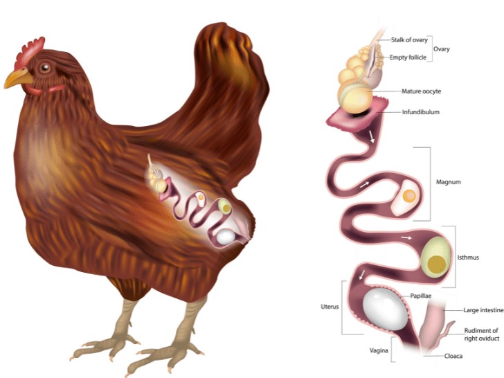
the mature yolk (ovum) waits here, wrapped in its follicle; on the LH surge the follicle ruptures at the stigma
↓ OVULATION — the yolk is released
the funnel actively engulfs the yolk. FERTILISATION happens here, within minutes, if sperm are present — it must occur before any albumen coats the ovum. The first (chalaziferous) albumen layer is added; the chalazae themselves form from it later, by twisting as the egg rotates through the magnum
↓ about 15 minutes spent here
glands pour out the thick albumen (egg white) around the yolk — the bulk of the egg white is laid down in this one part
↓ about 3 hours spent here
the inner and outer shell membranes are secreted around the albumen; the egg takes its final oval shape here. The egg still looks soft and wrinkled — it is not yet filled out
↓ about 1¼–1½ hours spent here
first, water and salts pass in through the membranes and 'plump' the egg to full size; then the shell is built — calcium carbonate (calcite crystals) deposited on the outer membrane, about 2 g of calcium per egg; shell pigment (in brown-egg breeds) and finally the cuticle are added at the end
↓ about 20–21 hours spent here — by far the longest stay
no part of the egg is made here. The sperm-storage tubules sit at its junction with the uterus (see the breeder section below). For laying, the egg turns so that it leaves blunt end first
↓ a few minutes only
the finished egg is laid. As it cools outside, the contents shrink slightly and the two shell membranes separate at the blunt end, forming the air cell
| Part | Length | Time Spent | What Is Added / What Happens |
|---|---|---|---|
| Infundibulum | ~9 cm | ~15 minutes | Yolk received; fertilisation (in breeders); chalaziferous albumen layer added (the chalazae form from it by twisting in the magnum) |
| Magnum | ~33 cm | ~3 hours | Albumen (egg white) — the bulk of it |
| Isthmus | ~10 cm | ~1¼–1½ hours | Inner and outer shell membranes; egg shape fixed |
| Uterus (shell gland) | ~11–12 cm | ~20–21 hours | Plumping (water + salts); shell (CaCO₃); pigment; cuticle |
| Vagina | ~8–12 cm | A few minutes | Nothing added; sperm storage at its upper end; egg turned blunt-end first and laid |
| Part of Egg | Formed In | Share of Egg Weight | Notes |
|---|---|---|---|
| Yolk | Ovary | About 31–33% | Made over days, not hours — the ovum plus its food store; carries the germinal disc |
| Albumen (white) | Magnum (main part); completed in uterus | About 57–60% | Four layers; the chalazae — twisted albumen cords — hold the yolk centred |
| Shell membranes | Isthmus | Part of the shell share | Two membranes; they separate at the blunt end to form the air cell after laying |
| Shell + cuticle | Uterus (shell gland) | About 9–12% | About 95% calcium carbonate; roughly 2 g calcium per shell; thousands of pores let the embryo breathe; cuticle seals them |
| Hormone / Signal | Source | Role in Egg Formation |
|---|---|---|
| Light (photoperiod) | Eyes → hypothalamus | Long days (about 14–16 hours of light for layers) switch on GnRH — light is the master switch of laying |
| FSH | Anterior pituitary | Grows the small follicles of the hierarchy |
| Oestrogen | Small growing follicles | Orders the liver to make yolk precursors; builds medullary bone — the hen's calcium store for shells |
| Progesterone | F1 (largest) follicle | Triggers the LH surge by positive feedback — a bird speciality |
| LH | Anterior pituitary | The surge ruptures the F1 follicle — ovulation |
| Prolactin | Anterior pituitary | Rises in broodiness — switches laying OFF and starts incubation behaviour |
Every reproductive event — puberty, the oestrous cycle, ovulation, pregnancy, parturition, spermatogenesis — is run by one command chain: the hypothalamus releases GnRH, the anterior pituitary answers with FSH and LH, and the gonads respond with the steroid hormones. Around this HPG axis (hypothalamus → pituitary → gonad) sit the supporting hormones: prostaglandin from the uterus, oxytocin, prolactin, inhibin, relaxin and melatonin. Learn the axis and the whole of reproduction makes sense.
| Hormone | Made By | Nature | Role in the Female | Role in the Male |
|---|---|---|---|---|
| GnRH | Hypothalamus (tonic + surge centres) | Decapeptide (10 amino acids) | Releases FSH and LH from the pituitary — the master switch of the cycle; the pre-ovulatory GnRH surge causes the LH surge | Releases FSH and LH in pulses (about 4–12 peaks a day) |
| FSH | Anterior pituitary (gonadotroph cells) | Glycoprotein | Grows the ovarian follicles; switches on aromatase in granulosa cells so oestradiol is made | Acts on Sertoli cells — supports spermatogenesis and makes inhibin and androgen-binding protein |
| LH (in the male also called ICSH) | Anterior pituitary | Glycoprotein | The surge ruptures the follicle (ovulation); then builds the corpus luteum and keeps it secreting progesterone | Acts on Leydig cells — orders testosterone production |
| Oestradiol (the main oestrogen) | Granulosa cells of the follicle; also placenta | Steroid (C18) | Brings the signs of heat (oestrus behaviour, tract changes, mucus); grows the duct system and mammary gland; feedback — negative most of the cycle, POSITIVE at high levels to trigger the LH surge | Needed in small amounts for libido and brain/behaviour development; excess suppresses the axis |
| Progesterone (P4) | Corpus luteum; also placenta (species vary) | Steroid (C21) | THE pregnancy hormone — prepares the endometrium, keeps the uterus quiet, blocks GnRH (negative feedback), grows the mammary gland | No primary role |
| Testosterone (the main androgen) | Leydig (interstitial) cells of the testis | Steroid | Present in small amounts as the raw material from which oestradiol is made | Spermatogenesis (with FSH), male duct system and accessory glands, secondary sex characters, libido and male behaviour |
| Inhibin | Granulosa cells (female); Sertoli cells (male) | Protein | Selectively holds back FSH release only — fine-tunes how many follicles grow | Selectively holds back FSH — the testis reporting 'enough' to the pituitary |
| Prostaglandin F2α (PGF2α) | Uterine endometrium (non-pregnant uterus) | Fatty-acid derivative | Destroys the corpus luteum (luteolysis) when there is no pregnancy — so the cycle starts again; also contracts the uterus | Also present in semen (made by the seminal vesicles) — helps sperm transport through the female tract |
| Oxytocin | Made in the hypothalamus, released from the posterior pituitary; also made by the CL in ruminants | Peptide (9 amino acids) | Contracts the uterus at parturition; causes milk let-down; in ruminants, luteal oxytocin drives the PGF2α pulses that end the cycle | Ejaculation — contracts the duct system |
| Prolactin | Anterior pituitary | Protein | Mammary development and milk production; in the BITCH it is the hormone that keeps the CL alive (luteotrophic); causes broodiness in birds | Minor role |
| Relaxin | Corpus luteum (sow); placenta (mare, bitch) | Polypeptide | Relaxes the pelvic ligaments and softens the cervix before birth; in the bitch it is used as a blood test for pregnancy (from about day 25) | — |
| Melatonin | Pineal gland (released at night) | Indoleamine | Reads day length and sets the breeding season — short-day breeders (ewe, doe) start cycling as nights lengthen; long-day breeders (mare) as days lengthen | Same seasonal role via the female axis |
| Activin | Granulosa cells (female); Sertoli cells (male) | Protein | Stimulates FSH release — the exact opposite of inhibin; supports follicle growth | Stimulates FSH release; local growth role inside the testis |
| AMH (anti-Müllerian hormone) | Sertoli cells of the FOETAL testis; granulosa cells of small follicles in the adult female | Glycoprotein | In the adult it mirrors the ovarian reserve; a blood AMH test detects an ovarian remnant in a spayed bitch or queen | In the foetus it makes the Müllerian ducts regress — so the male develops no uterus or tubes; in the adult, a blood test proves a retained (cryptorchid) testicle is present |
| Kisspeptin | Hypothalamic neurones (KISS1 neurones) | Peptide | The upstream trigger of GnRH — it starts puberty and drives the GnRH surge before ovulation | The upstream trigger of the GnRH pulses that run the testis |
| Placental lactogen | Placenta (best known in ruminants) | Protein | Develops the mammary gland during pregnancy and prepares it for milk production | — |
Oestradiol is made by two cells working as a team, each driven by a different gonadotrophin. Neither cell can finish the job alone:
| Cell | Driven By | What It Does |
|---|---|---|
| Theca interna (follicle wall) | LH | Cholesterol → androgens (testosterone) — the raw material |
| Granulosa (inside the follicle) | FSH | Takes the androgens and, using the enzyme AROMATASE, converts them → oestradiol |
| Leydig cell (testis) | LH | Cholesterol → testosterone |
| Sertoli cell (testis) | FSH | Converts some testosterone → oestradiol; also makes inhibin and androgen-binding protein |
One line to remember: Cholesterol → Testosterone → Oestradiol, and aromatase is the final switch.
| Hormone | Species | Why It Is Special |
|---|---|---|
| eCG (equine chorionic gonadotrophin, old name PMSG) | Mare | Made by the endometrial cups of the pregnant mare (about day 40–120 of pregnancy); in the mare it binds LH receptors only (LH-like, luteotrophic) — it ovulates and luteinises existing follicles to form the accessory corpora lutea of early pregnancy; its FSH-like action appears only in other species |
| Relaxin | Bitch | The basis of the commercial blood test for pregnancy in dogs — detectable from about day 22–30 after mating |
| Prolactin | Bitch | The luteotrophic hormone of the dog — without it the CL fails; blocking prolactin (with cabergoline) therefore ends pseudopregnancy and can end pregnancy |
| Progesterone from the placenta | Mare, ewe | In the mare the placenta takes over progesterone-type support after about day 100 (as progestagens); in the ewe the placenta takes over in the second half — in the cow and doe the CL is needed for the whole pregnancy |
| Melatonin | Ewe, doe, mare | The season setter — sheep and goats breed as days shorten (autumn), the mare as days lengthen (spring); the cat is also a long-day breeder |
Most of the time the system runs on NEGATIVE feedback: progesterone (and moderate oestradiol) hold GnRH, FSH and LH down, which is why nothing ovulates during the luteal phase or pregnancy. The exception is the end of the follicular phase, when oestradiol from a mature follicle rises high and switches to POSITIVE feedback — the hypothalamus answers with a GnRH surge, the pituitary with an LH surge, and the follicle ruptures (in the cow, ovulation follows about 24–32 hours after the ONSET OF OESTRUS — about 21–30 hours after the LH surge begins). Inhibin fine-tunes FSH alone. When there is no pregnancy, the uterus speaks last: endometrial PGF2α kills the CL, progesterone falls, the brake comes off, and a new cycle begins.
Almost every hormone of the previous topic is also sold as a medicine. On farms these products are big business: they let the farmer breed a whole group on one day (synchronisation), fix problem breeders, superovulate valuable donors for embryo transfer, and time parturition. This topic lists what is used, in which sex, in which species — and why.
| Hormone / Preparation | Main Commercial & Clinical Uses | Species |
|---|---|---|
| GnRH (gonadorelin, buserelin — e.g. Receptal) | Ovsynch fixed-time AI programme; treatment of follicular cysts; ovulation induction; given at insemination for delayed ovulation | Cattle mainly; also mare; in the bitch, oestrus induction is by the deslorelin implant form (see the deslorelin row), not plain GnRH injections |
| PGF2α (dinoprost 25 mg IM; analogue cloprostenol 500 µg IM) | Oestrus synchronisation (works only when a functional CL is present, from about day 5 of the cycle); open pyometra; mummified foetus; persistent CL; luteal cysts; induction of abortion and of parturition | Cattle, buffalo, small ruminants, mare, sow |
| Progesterone devices — CIDR (1.38–1.9 g) and PRID (1.55 g) intravaginal | Oestrus synchronisation in cycling and anoestrous females — the device acts as an artificial CL; removed after 7–12 days, heat follows | Cattle, buffalo, sheep, goat |
| Progestogen sponges (vaginal) + eCG at removal | Out-of-season breeding and synchronisation — the classic small-ruminant programme | Sheep, goat |
| MGA (melengestrol acetate) — in feed | Suppresses oestrus in feedlot heifers while improving weight gain — a production use, not a breeding one | Beef heifers |
| Altrenogest (oral progestogen) | Suppresses and synchronises oestrus — daily feeding, heat 3–7 days after withdrawal | Mare; also gilts |
| FSH (e.g. Folltropin) — injections every 12 hours for 4–5 days | SUPEROVULATION of donor cows for embryo transfer (prostaglandin given on day 3–4) | Cattle (embryo transfer) |
| eCG / PMSG (single injection) | Superovulation (cheaper single-dose alternative); follicle growth in anoestrous ewes/does at sponge removal; oestrus induction | Cattle, sheep, goat, pig |
| hCG (LH-like; e.g. Chorulon) | Ovulation induction — the mare with a mature follicle (ovulation in about 36–48 hours); cystic ovaries in cattle (label doses: 3,000 IU IV or 10,000 IU IM); delayed ovulation at AI (1,000–1,500 IU) | Mare, cattle |
| Oxytocin | Milk let-down; uterine inertia at parturition (only when the cervix is open); helps uterine involution and retained-placenta management | All farm species, bitch |
| Melatonin implants | Advance the breeding season — implanted before the season so ewes/does cycle earlier | Sheep, goat |
| Deslorelin implant (GnRH agonist) | Induces oestrus and ovulation in the anoestrous bitch; in the mare induces ovulation of a mature follicle | Bitch, mare |
| Programme | How It Runs | Used In |
|---|---|---|
| Ovsynch | Day 0 GnRH → Day 7 PGF2α → Day 9 second GnRH → fixed-time AI 0–20 hours later, no heat detection needed | Dairy cattle |
| CIDR programme | CIDR in for 7 days (+ GnRH at insertion in many versions) → remove CIDR + PGF2α → heat in 2–3 days, AI on observed heat or fixed time | Cattle, buffalo |
| Double prostaglandin | Two PGF2α injections 11–14 days apart — after the second, the whole group is at the same cycle stage; breed on heat or fixed time | Cattle, buffalo, mare |
| Sponge + eCG | Progestogen sponge 12–14 days → remove + eCG injection → fixed-time AI about 48–55 hours later in ewes (43–46 hours in does); works even outside the breeding season | Sheep, goat |
| Hormone / Preparation | Use | Species / Notes |
|---|---|---|
| hCG stimulation test | DIAGNOSIS of a retained testicle: hCG is injected and testosterone measured before/after — a rise proves testicular tissue is present | Stallion, dog (cryptorchid testing) |
| GnRH / hCG | Treatment attempts in some cryptorchid young animals (success is limited — surgery remains the standard); GnRH also used to test pituitary function | Dog, farm species |
| Testosterone and anabolic steroids | Occasionally used for depressed libido in breeding males; mainly of historical/limited use — excess SUPPRESSES spermatogenesis by feedback | Breeding males — use with caution |
| Deslorelin implant (Suprelorin) | REVERSIBLE chemical castration — months of suppressed testosterone and fertility without surgery; also used for hormone-driven problems such as adrenal disease in ferrets | Dog (male and female contraception) |
| Growth-promoting implants — zeranol; trenbolone acetate + oestradiol | Placed under the ear skin to increase growth rate and feed efficiency in beef production — a purely commercial use of sex hormones; banned in the European Union | Beef steers and heifers |
Most hormone medicines are not the natural hormone at all — they are AGONISTS: man-made copies built to last longer or act stronger than the original. The natural hormones are often destroyed in minutes (GnRH lasts only a few minutes), so the copy is what reaches the syringe.
| Natural Hormone | Analogue / Agonist Preparations | Why the Copy Is Preferred |
|---|---|---|
| GnRH | Gonadorelin, buserelin, fertirelin | Stronger and longer-acting than natural GnRH; reliable LH release from one injection |
| GnRH (long-acting) | Deslorelin implant | One implant works for months — first stimulates (flare), then switches the axis OFF by down-regulation; the basis of reversible contraception in the dog |
| PGF2α | Cloprostenol, luprostiol | About 200 times more potent than natural dinoprost — a 500 µg dose does the work of 25 mg, with fewer side effects |
| Progesterone | Altrenogest, melengestrol acetate (MGA), medroxyprogesterone, proligestone | Can be given by mouth or in feed and last far longer than natural progesterone, which needs oily depot injections or devices (CIDR/PRID) |
| LH | hCG (borrowed from human pregnancy urine/placenta) | A ready-made LH-like hormone with a much longer half-life than LH itself |
| FSH | eCG / PMSG (from pregnant mare serum) | A ready-made FSH-like hormone in a single injection, instead of twice-daily FSH for 4–5 days |
| Oxytocin | Carbetocin | Long-acting oxytocin copy — one injection covers the whole of farrowing/parturition instead of repeated short doses |
ANTAGONISTS do the opposite job: they block a hormone's receptor or switch off its release. They are how the veterinarian stops a pregnancy, dries up false pregnancy, or shuts down an over-active axis.
| Blocker | What It Blocks | Veterinary Use |
|---|---|---|
| Aglepristone (Alizin) | Progesterone receptors | THE standard drug to end an unwanted pregnancy in the bitch (usable up to about day 45) — it removes progesterone's support without needing progesterone to fall |
| Mifepristone | Progesterone receptors | Antiprogestin used in some pregnancy-termination and pyometra protocols |
| Cabergoline | Prolactin (it is a dopamine agonist — dopamine is prolactin's natural brake) | False pregnancy (pseudopregnancy) in the bitch; suppresses unwanted lactation; used in pregnancy termination after about day 30, when the CL depends on prolactin |
| Bromocriptine | Prolactin (dopamine agonist) | Older drug for the same jobs as cabergoline — more vomiting, so largely replaced by it |
| GnRH antagonists (cetrorelix, ganirelix) | GnRH receptors — immediate blockade, no flare first | Limited veterinary use so far (research and special protocols) — unlike deslorelin, they stop LH/FSH at once |
| Clomiphene | Oestrogen feedback at the hypothalamus | Blocks oestrogen's negative feedback, so GnRH and gonadotrophins rise — an ovulation inducer borrowed from human medicine — but NOT established in veterinary practice: it failed to induce fertile oestrus in bitches and acts agonist-like in dogs |
| Tamoxifen | Oestrogen receptors (a SERM — blocker in some tissues, weak agonist in others) | Occasionally used around mammary tumours; in the bitch it can itself cause heat signs and pyometra — use with great care |
| Osaterone acetate; delmadinone acetate; finasteride | Androgen action (receptor blockers / 5α-reductase inhibitor) | Benign prostatic hyperplasia (BPH) and perianal adenoma in the older male dog — they shrink the prostate without castration |
| Atosiban | Oxytocin receptors | A womb-relaxing (tocolytic) drug of human medicine; not established in routine veterinary practice |
The pair to remember for the exam: deslorelin is an agonist that ends up behaving like an antagonist (down-regulation), while aglepristone and cabergoline are the two true blockers a small-animal vet uses every week — one blocks progesterone, the other removes prolactin.
The oestrous cycle is the female's repeating rhythm of fertility, run by the hormones of the earlier topic: follicles grow under FSH, oestradiol brings heat, the LH surge ovulates, progesterone holds the luteal phase, and PGF2α resets the clock. The cycle is divided into four stages — two follicular (proestrus, oestrus) and two luteal (metestrus, dioestrus).
| Stage | What Happens in the Ovary | Hormone in Charge | What the Farmer Sees / Tract Changes |
|---|---|---|---|
| Proestrus | A dominant follicle grows and matures | Oestradiol rising | Tract prepares: vulva swells, mucus begins; in the BITCH this is the stage of bloody discharge (the bleeding is proestrus, NOT heat itself); female attracts males but refuses mating |
| Oestrus (heat) | Follicle matures; LH surge; ovulation (timing varies — Table 2) | Oestradiol at peak | Standing heat — the female accepts the male; clear stretchy mucus in cattle; frequent urination and 'winking' in the mare; restlessness, bellowing, milk drop in the cow |
| Metestrus | The ruptured follicle becomes a corpus haemorrhagicum, then a young CL | Progesterone rising | Heat signs fade; some heifers show metestrus bleeding (a little blood on the tail 1–2 days after heat — normal, from oestrogen withdrawal) |
| Dioestrus | CL fully functional; uterus ready for pregnancy | Progesterone dominant | Quiet female; if no pregnancy, the uterus releases PGF2α at the end of this stage, the CL dies, and a new cycle begins |
| Species | Cycle Length | Heat Duration | Ovulation — When | Pattern |
|---|---|---|---|---|
| Cow | About 21 days (18–24) | 12–18 hours | 12–14 hours AFTER heat ends | Polyestrus — cycles all year |
| Buffalo | About 21 days | 12–24 hours | Towards the end of / just after heat | Polyestrus, but silent heat is very common and breeding is seasonal in practice (cooler months) |
| Mare | 21–22 days | 5–7 days (long!) | 1–2 days BEFORE heat ends | Seasonally polyestrus — long-day breeder (spring/summer) |
| Ewe | 16–17 days | 24–36 hours | Towards the end of heat | Seasonally polyestrus — short-day breeder (autumn) |
| Doe (goat) | About 21 days | 24–48 hours | Towards the end of heat | Seasonally polyestrus — short-day breeder |
| Sow | About 21 days | 2–3 days | About 40 hours after heat starts (last third of heat) | Polyestrus — cycles all year |
| Bitch | One cycle every 6–7 months | Heat (oestrus) 5–9 days, after 7–10 days of proestrus bleeding | About 2 days after the LH surge — and the eggs are still immature, needing 2–3 more days | Monoestrus — one cycle per season; long dioestrus follows whether pregnant or not (hence false pregnancy) |
| Queen (cat) | 14–21 days in season | 5–7 days | Only after mating — INDUCED ovulator | Seasonally polyestrus — long-day breeder |
Semen is spermatozoa plus seminal plasma (from the accessory glands). Evaluating it is the core of the male breeding-soundness examination: a male is only as good as his ejaculate. The species differ enormously — the boar gives a cupful of dilute semen, the ram a teaspoon of concentrate — because their accessory glands differ.
| Species | Volume per Ejaculate | Concentration (per mL) | Notes |
|---|---|---|---|
| Bull | About 4 mL (1–15) | About 1,000–2,000 million | Creamy white, opaque; pH about 6.7 |
| Buffalo bull | About 3–5 mL | About 600–1,200 million | Similar to the bull, slightly lower volume |
| Stallion | About 70 mL (30–250) | About 100–300 million | Has a gel fraction (from the seminal vesicles) that is filtered out; pH about 7.4 |
| Boar | About 250 mL (125–500) — the largest | About 200–300 million | Fractionated ejaculate with a large gel (coagulum) portion; pH about 7.4 |
| Ram | About 1 mL (0.7–3) | About 2,000–3,000 million — the most concentrated | Thick and creamy; sperm form over 30% of the ejaculate (spermatocrit) |
| Buck | About 1 mL | About 2,000–3,000 million | Like the ram — small volume, very concentrated |
| Dog | About 10 mL (1–25), in 3 fractions | About 100–500 million | Only the second fraction is sperm-rich; dog semen contains NO fructose |
| Cock | About 0.5–1 mL | About 3,000–5,000 million | Collected by abdominal massage for poultry AI |
Pregnancy diagnosis answers the farm's most valuable question as early and as safely as possible: is she pregnant, and if not, get her bred again without wasting a cycle. Every method has its earliest reliable day — too early means false negatives, and rough palpation too early can damage the pregnancy itself.
| Species | Method of Choice | Earliest Reliable Day | Special / Laboratory Options |
|---|---|---|---|
| Cow | Rectal palpation; transrectal ultrasound | Ultrasound: day 26–28 (heartbeat); Palpation: day 35 — membrane slip about day 33, amniotic vesicle day 30–50, placentomes from about day 75–90 | Milk/plasma progesterone day 21–24 (LOW rules pregnancy OUT; high does not prove it); pregnancy-associated glycoproteins (PAGs) from about day 28 |
| Buffalo | Rectal palpation; ultrasound | Ultrasound day 28–30; palpation usually day 45–60 (deeper pelvis, later signs) | Progesterone assay as in cattle |
| Mare | Transrectal ultrasound | Day 12–14 — the vesicle; recheck day 14–16 to find and crush a twin; heartbeat about day 25 | Blood eCG day 40–120 (false positive if the foetus dies after cups form); Cuboni test (oestrogens in urine) about day 120–300; oestrone sulphate later |
| Ewe / Doe | Transabdominal ultrasound | Day 30–40 (transrectal scanning works earlier, about day 25) | Radiography in late pregnancy for numbers; PSPB/PAG blood tests |
| Sow | Ultrasound (Doppler / A-mode / real-time) | Day 21–24 by real-time scanning | Non-return to heat at day 18–24 is the everyday herd check |
| Bitch | Abdominal palpation; ultrasound; relaxin test | Palpation: day 21–30 (individual tense swellings — the window closes after about day 35); Ultrasound: best day 25–35; Relaxin blood test: day 22–30 | Radiography from day 45 (skeletons mineralise) — the method for COUNTING foetuses, best after day 55 |
| Queen | Ultrasound; palpation | Ultrasound day 20–25; relaxin about day 25–30 | Radiography from about day 40 |
| Camel | Behaviour + rectal palpation | 'Cocking of the tail' when approached by a male/person from day 14–15 — about 95% reliable in quiet females | Ultrasound as in cattle |
The placenta is the foetus's life-support organ — lungs, gut, kidney and endocrine gland in one. Veterinary exams test it two ways: by the SHAPE of the attachment (gross type) and by HOW MANY tissue layers separate foetal from maternal blood (histological type). The layer count decides a life-or-death practical point: whether antibodies can cross before birth.
| Gross Type | Species | How It Looks |
|---|---|---|
| Diffuse, complete | Mare | Villi cover the whole chorion (as tiny microcotyledons) — attachment everywhere; the placenta comes away cleanly and completely |
| Diffuse, incomplete (folded) | Sow | Villi over most of the chorion but absent at the ends and over folds |
| Cotyledonary | Cow, buffalo, sheep, goat | Attachment only at placentomes — a foetal cotyledon locking into a maternal caruncle like a button; cow about 75–120 placentomes, sheep and goat about 90–100 |
| Zonary | Bitch, queen | A belt (zone) of attachment around the middle of the chorion; the edge has a marginal haematoma — the source of the green pigment (uteroverdin) seen in the bitch's discharge at whelping |
| Discoid | Primates, rodents (for comparison) | A single disc of attachment — not found in farm animals |
| Type | Layers | Species | Antibodies Cross Before Birth? |
|---|---|---|---|
| Epitheliochorial | 6 — the full set, both epithelia intact | Mare, sow | NO — the newborn is born without antibodies; colostrum is life or death |
| Synepitheliochorial | 6, but special binucleate cells of the chorion fuse with the uterine epithelium | Cow, buffalo, sheep, goat (ruminants) | NO — colostrum is essential in calves, lambs and kids too |
| Endotheliochorial | 4 — the uterine epithelium and stroma are eroded away | Bitch, queen | A small part crosses (about 5–10%) — most still comes from colostrum |
| Haemochorial | 3 — foetal tissue bathes directly in maternal blood | Primates, rodents (for comparison) | YES — freely; their newborns are born already protected |
Parturition is started by the foetus itself. When the foetus is mature, its own stress axis fires the starting gun — one of the most elegant mechanisms in physiology, and a favourite exam topic. Everything after that is the mother's body answering.
| Stage | What Happens | Cow / Buffalo | Mare | Ewe / Doe | Sow | Bitch |
|---|---|---|---|---|---|---|
| Stage 1 — dilation | Cervix opens; uterus contracts; foetus rotates into position; restlessness, isolation, colic-like signs | 2–6 hours | 1–4 hours | 2–6 hours | 2–12 hours | 6–12 hours (nesting, temperature drop about 1 °C a day before) |
| Stage 2 — expulsion | Abdominal straining delivers the foetus (water bag breaks first) | 30 minutes–2 hours (longer in heifers) | 15–30 minutes — FAST; longer is an emergency | 30–60 minutes per lamb/kid | 15–30 minutes between piglets | 5–60 minutes between pups |
| Stage 3 — placenta | Placenta separates and is expelled | Up to 8–12 hours; RETAINED if over 24 hours | 1–3 hours; RETAINED if over 3 hours — an emergency (laminitis risk) | 2–3 hours | During farrowing or within about 1–4 hours after the last piglet | With each pup or in pairs |
| Term | Meaning | The Normal at Birth |
|---|---|---|
| Presentation | Which part of the foetus enters the pelvis first, and the direction of its spine | Longitudinal ANTERIOR (head and forelegs first) in most species; longitudinal POSTERIOR is also normal in litter-bearing species and acceptable in calves |
| Position | Where the foetus's back lies relative to the mother's pelvis | Dorso-sacral — the foetus's back towards the mother's sacrum (foetus 'right way up') |
| Posture | How the head and limbs are arranged | Head resting on extended forelegs (the 'diving' posture); any flexion — head back, leg back — is a dystocia cause |
At every birth, the arrangement of the fetus inside the dam — its DISPOSITION — is described by three words, always in this order: presentation, position and posture. Any fault in them is called MALDISPOSITION, and it is a fetal cause of dystocia (difficult birth).
| Term | What it describes | How it is judged |
|---|---|---|
| Presentation | The relation of the long axis (spinal axis) of the fetus to the long axis of the dam — i.e. WHICH PART of the fetus faces the birth canal | Longitudinal (anterior or posterior), transverse or vertical |
| Position | The relation of the DORSUM (back) of the fetus — in transverse presentation, of the HEAD — to the four quadrants of the mother’s pelvis | Quadrants: sacrum (top), pubis (floor), right ilium, left ilium |
| Posture | The relation of the MOVABLE PARTS of the fetus — head, neck and limbs — to the fetus’s OWN body | Each part extended (normal) or flexed / deviated (abnormal) |
| Presentation | What it means | Normal or abnormal |
|---|---|---|
| Anterior (cranial) longitudinal | Head and forelimbs enter the pelvis first; fetal spine parallel to the dam’s spine | NORMAL — about 95% of calves, and the usual presentation in mare, ewe and doe |
| Posterior (caudal) longitudinal | Hindquarters and hind limbs enter the pelvis first; fetal spine parallel to the dam’s spine | NORMAL — about 5% of calves; in sow, bitch and queen up to 40–45% of fetuses are born this way, normally |
| Transverse dorsal | Fetal spine lies ACROSS the dam’s spine and the fetus’s BACK faces the pelvic opening | ABNORMAL — the fetus cannot enter the canal; rare, worst in the mare |
| Transverse ventral | Fetal spine lies across the dam’s spine and the BELLY (with all four limbs) faces the pelvic opening | ABNORMAL — limbs may enter the canal but delivery is impossible without correction |
| Vertical / oblique | Fetus sits upright or slanting in the uterus | ABNORMAL — rare |
| Position | What it means | Status |
|---|---|---|
| Dorso-sacral | The fetus’s back lies against the mother’s SACRUM (the roof of the pelvis) | NORMAL — the only position for an easy normal birth in large animals |
| Right dorso-ileal | The fetus’s back lies against the mother’s RIGHT ilium (right wall of the pelvis) | Abnormal — fetus rolled about a quarter-turn; usually corrected by rotation |
| Left dorso-ileal | The fetus’s back lies against the mother’s LEFT ilium (left wall of the pelvis) | Abnormal — corrected by rotation |
| Dorso-pubic | The fetus’s back lies against the PUBIS (the floor of the pelvis) — the fetus is upside-down — also called the VENTRAL position | Abnormal — the most unfavourable position; the soles of the feet point upward |
| Cephalo-ileal (right or left) | Used only in TRANSVERSE presentation: the fetus’s HEAD lies toward the mother’s right or left ilium | Abnormal (belongs to transverse presentation) |
NORMAL POSTURE (anterior presentation): head and neck fully EXTENDED, the chin resting on the knees and the muzzle reaching the fetlocks, both forelimbs extended straight into the canal — the “diving” posture. In posterior presentation, both hind limbs are fully extended behind the fetus into the canal.
| Abnormal posture | What exactly happens | Seen in |
|---|---|---|
| Lateral deviation of head and neck | Head is bent sideways, to the right or left, against the fetus’s own chest/flank | Anterior presentation — a very common cause of dystocia in calf and lamb |
| Ventral (downward) deviation of head | Head is bent DOWN between the forelimbs | Anterior presentation |
| Vertex posture | Head flexes at the poll so the FOREHEAD (vertex) faces the pelvis and the nose points down/back | Anterior presentation — mostly the foal |
| Complete downward deviation of head (nape presentation) | The head is bent fully DOWN until the chin touches the breast and the NAPE of the neck faces the pelvis | Anterior presentation |
| Upward deviation of head | Head is thrown UP and back over the fetus’s own back | Anterior presentation — rare |
| Fetlock flexion | One or both forelimbs bent at the FETLOCK joint | Anterior presentation |
| Carpal (knee) flexion | One or both forelimbs bent at the CARPUS, the folded knee blocking the pelvis | Anterior presentation — common |
| Incomplete extension of the elbow | The forelimb is not fully straightened — it stays partly bent at the ELBOW joint, so the limb cannot enter the canal properly | Anterior presentation |
| Shoulder flexion (retained forelimb) | The whole forelimb lies folded back ALONG THE BODY; only the shoulder reaches the pelvis | Anterior presentation |
| Foot-nape posture | A forefoot rests on top of the fetus’s neck/nape and can tear the roof of the vagina | Anterior presentation — a mare problem |
| Hock flexion | In posterior presentation, one or both hind limbs are bent at the HOCK | Posterior presentation |
| Hip flexion — BREECH | Both hind limbs fold forward UNDER the body at the hip; only the tail and rump enter the pelvis | Posterior presentation — this is the true BREECH |
| Dog-sitting posture | In ANTERIOR presentation the hind limbs are pushed forward under the body, so the fetus “sits” like a dog | Almost only the foal |
Some faults are so common that obstetricians give the whole combination a single name. These names are exam favourites — each name must be broken into its presentation + position + posture:
| Named disposition | What it actually is (presentation + position + posture) | Species / notes |
|---|---|---|
| Breech presentation | Posterior longitudinal presentation + dorso-sacral position + BOTH HIPS FLEXED — hind limbs folded forward under the body, only the tail and rump at the pelvis | Calf, lamb, puppy — the classic named abnormality |
| Butt presentation | Anterior longitudinal presentation + BOTH SHOULDERS FLEXED — both forelimbs folded back, so the chest/butt of the fetus faces the pelvis | Calf, lamb, foal |
| Dog-sitting presentation | Anterior longitudinal presentation, but BOTH HIND LIMBS pushed forward under the body — the fetus sits like a dog | Almost only the FOAL |
| Vertex (poll) posture | Anterior presentation with the head flexed at the POLL, so the forehead (vertex) faces the pelvis | Mostly the foal |
| Foot-nape posture | Anterior presentation with a FOREFOOT resting on the nape of the neck — the foot can tear the roof of the vagina | MARE |
| “Head back” (field name) | Anterior presentation with LATERAL DEVIATION of the head and neck — the head lies back against the fetus’s own side | Calf and lamb — among the commonest dystocias |
| “Leg back” (field name) | Anterior presentation with SHOULDER FLEXION — the whole forelimb folded back along the body | Calf, lamb, foal |
| “Knee up” / knee-flexed (field name) | Anterior presentation with CARPAL FLEXION — the folded knee blocks the pelvic inlet | Calf, lamb, foal |
| “Upside-down calf” (field name) | DORSO-PUBIC position — the fetus lies upside-down, soles of the feet pointing up | Calf |
| Cross presentation (field name) | TRANSVERSE presentation — the fetus lies across the dam’s spine (dorsal or ventral form) | Mare and cow — rare |
| Nape presentation (head-breast posture) | Anterior presentation with COMPLETE downward deviation of the head — chin tucked to the breast, nape facing the pelvis (the incomplete form is the vertex posture) | Calf, foal |
| Species | Normal presentation | Normal position | Normal posture |
|---|---|---|---|
| Cow & buffalo | Anterior longitudinal (about 95%); posterior longitudinal in about 5% | Dorso-sacral | Head, neck and forelimbs extended (posterior: hind limbs extended) |
| Mare | Anterior longitudinal | Dorso-sacral | Head, neck and forelimbs extended |
| Ewe & doe | Anterior longitudinal (posterior also common in twins) | Dorso-sacral | Head resting on the extended forelimbs |
| Sow | Anterior AND posterior — both normal (up to 40–45% posterior) | Dorso-sacral or dorso-ileal may pass | Forelimbs folded back in anterior; hind limbs extended in posterior |
| Bitch & queen | Anterior about 60%, posterior about 40% — both normal | Dorso-sacral | Forelimbs extended in anterior presentation |
| Term | Meaning |
|---|---|
| Cattle plaque | Rinderpest |
| Equine plaque | African horse sickness |
| Goat plaque | PPR |
| Fowl plaque | Avian influenza |
| Cat plaque | Feline panleukopenia |
| Rabbit plaque | Tularemia — Francisella tularensis |
| Duck plaque | Duck viral enteritis |
| Bubonic plaque | Yersinia pestis |
| Lung plaque | CBPP |
| White plaque | Tuberculosis — Mycobacterium bovis in animals |
| Term | Meaning |
|---|---|
| Black quarter / Blackleg | Clostridium chauvoei |
| Blackhead | Histomonas meleagridis |
| Black disease | Clostridium novyi Type B |
| Black flies | Simulium spp. (family Simuliidae) |
| Black death | Yersinia pestis |
| Black tongue | Niacin deficiency |
| Term | Meaning |
|---|---|
| Pseudo FMD | Bluetongue |
| Pseudo cowpox | Milker's nodule |
| Pseudo rinderpest | PPR |
| Pseudo tuberculosis | Yersinia pseudotuberculosis |
| Pseudo rabies | Aujeszky's disease / infectious bulbar paralysis |
| Pseudo fowl plaque | Newcastle disease |
| Pseudo glanders | Equine epizootic lymphangitis |
| Term | Meaning |
|---|---|
| Bollinger bodies | Fowl pox |
| Joest bodies | Borna disease |
| Döhle's bodies | Aggregates of ribosomes in neutrophils |
| Koch blue bodies | Theileriosis |
| Councilman bodies | Yellow fever in man |
| Negri bodies | Rabies |
| Guarnieri bodies | Smallpox (variola) and vaccinia — all orthopoxviruses |
Labelled diagram of the hen's oviduct; labels in flow order:
| Stage / Part | Diagram labels |
|---|---|
| Follicles | Ovulated follicle |
| Infundibulum | 0.25 hour · Fertilization · Chalazae |
| Magnum | 3 hours · Albumen |
| Isthmus | 1.25 hours · Soft Shell |
| Uterus / Shell Gland | 21 hours · Hard Shell |
| Vagina | Seal pores · Sperm storage |
| Part | Site | Time |
|---|---|---|
| Yolk | Ovary | 7–9 days |
| Thick mucin | Infundibulum | 15–30 minutes |
| Albumin | Magnum | 2–3 hours |
| Shell membrane | Isthmus | 1.5 hours |
| Watery solution (plumping) | Shell gland | 3–5 hours (part of the shell-gland stay) |
| Shell | Shell gland | About 20–21 hours total stay (including plumping) |
| Bloom (mucus) | Shell gland (uterus) | 1–10 minutes |
| Nutrients | Milk | Egg |
|---|---|---|
| Water (gm) | 87 | 75 |
| Protein (gm) | 3.2 | 12 |
| Fat (gm) | 3.5 | 10 |
| Calcium (mg) | 118 | 60 |
| Phosphorus (mg) | 93 | 220 |
| Iron (mg) | Trace | 2.1 |
| Vit. A (IU) | 140 | 480–540 |
| Riboflavin (mg) | 0.17 | 0.45 |
| Niacin (mg) | 0.11 | 0.07 |
| Egg Part | Formation Process | Body System Involved | Approximate Duration |
|---|---|---|---|
| Yolk | Formed in the ovary from developing ova (follicles) | Reproductive (ovary) | 7–10 days |
| Yolk Release | Mature yolk released into the oviduct | Reproductive | Instant |
| Fertilization | Occurs in infundibulum | Reproductive | 15–30 minutes |
| Egg White (Albumen) | Albumen added around yolk | Reproductive (magnum) | 3 hours |
| Shell Membranes | Two membranes form around albumen | Reproductive (isthmus) | ~1.5 hours |
| Shell Formation | Calcium carbonate shell deposited | Reproductive (uterus/shell gland) | 20 hours |
| Cuticle (Bloom) | Protective coating added | Reproductive | Last hour |
| Laying | Egg exits the body | Reproductive | Instant |
What is Hay? Hay is grass, legumes, or other plant sources that have been cut, dried, and preserved in such a way that its nutrients are preserved with minimum loss and made available to livestock as feed during times of scarcity.
Color of good Hay: The color of the good-quality hay should be green; it should not be too coarse and include plant heads, leaves, and stems.
Characteristics of bad quality hay: Poor-quality hay is dry, bleached out, and coarse-stemmed. A dried, bleached, or coarse bale is still edible and provides some nutritional value as long as it is dry and not moldy, dusty, or rotting.
Crude Protein values of Hay: Lucerne Hay — 20.5% · Berseem Hay — 15.1% · Oat Hay — 10%
Steps in Hay Making: Cutting/Mowing · Tedding — spread fodder for faster drying · Raking — gathering the dried forage into windrows for baling · Baling — making bundles of hay · Stacking/storage
Best Suited Crops for Hay Making: Berseem · Lucerne · Oats
What is Silage? Silage is fermented, high-moisture-stored fodder that can be fed to ruminants such as cattle, buffalo, etc. Silage is the better way to conserve forage crops. A forage crop can be cut early and should have 30–35% dry matter to be ensiled successfully.
Fermentation (48 hours – 2 weeks): Silage undergoes anaerobic fermentation which starts about 48 hours after the silo is filled, and converts sugars to acids. Fermentation is essentially complete after about two weeks. Before anaerobic fermentation starts, there is an aerobic phase in which the trapped oxygen is consumed. When closely packed, the supply of oxygen is limited; and the acid fermentation brings about the decomposition of the carbohydrates present into acetic, butyric, and lactic acids.
If the fermentation process is poorly managed, silage acquires an unpleasant odor due to excess production of ammonia or butyric acid (like rancid butter). Silage inoculants are used to enhance fermentation. These contain one or more strains of lactic acid bacteria, and the most common is Lactobacillus plantarum. Other bacteria used in inoculants include Lactobacillus buchneri and Enterococcus faecium species.
Types of Silos: Bag Silo · Tower Silo · Trench Silo · Bunker Silo
Estimating Moisture Contents: The approximate moisture level of chopped silage can be determined using a "grab test." Squeeze the chopped forage tightly into a ball for 20 to 30 seconds, and then release quickly. Forage chopped into 3/8 to 2 inch pieces should be used for this test.
Hay and silage are the two ways of preserving green fodder for the lean season: hay preserves it by DRYING, silage by FERMENTATION. (Table below verified from university extension and FAO forage sources.)
| Point of difference | Hay | Silage |
|---|---|---|
| What it is | Forage cut and dried in sun/air and stored dry | Green forage chopped, packed airtight and preserved by fermentation |
| Preserving principle | Water is removed — too dry for moulds and bacteria to grow | Lactic-acid bacteria ferment plant sugars to lactic acid; the low pH stops spoilage |
| Moisture at storage | About 10–15% | About 60–70% |
| Dry matter (DM) | About 85–90% | About 30–40% |
| pH | Not applicable — a dry product | 3.8–4.5 in well-fermented silage (3.8–4.2 is best) |
| Suitable crops | Thin-stemmed, leafy crops that dry evenly — lucerne, berseem, oats and grasses | Thick-stemmed, sugar/starch-rich crops — maize (best), sorghum (jowar), bajra |
| Harvest stage | Grasses at flowering; legumes at about 1/3 to 1/2 bloom | Maize at the milk-to-dough stage (kernel milk line 1/2 to 2/3); sorghum at milk–dough stage |
| Time and weather needed | Needs several days of good sunshine for field curing | Chopped, packed and sealed the same day; ready to feed after about 3–6 weeks of fermentation |
| Storage | Bales or stacks under a roof or shed — no sealing needed | Airtight pit/bunker silo, silo bags or plastic-wrapped bales — must stay sealed |
| Nutrient losses | Leaf shattering and sun-bleaching (carotene/vitamin A falls); rain during curing spoils it | Fermentation and effluent (seepage) losses; keeps its green colour and carotene better |
| Main risks | Mould and heating — even spontaneous fire — if stored too wet; dustiness | Listeriosis and botulism in badly made silage; spoils fast once the silo is opened to air |
| Transport and trade | Light and dry — easy to transport, store and sell | Heavy because it is mostly water — costly to transport; usually fed near the silo |
Note: Best hay is prepared with Oat (Avena sativa). Best legume hay is prepared with Lucerne (alfalfa). Maize is the best silage crop.
| Type | Name | Mechanism | Key Immune Components | Examples | Onset (initiation time) |
|---|---|---|---|---|---|
| Type I | Immediate (Anaphylactic) | IgE-mediated; mast cell degranulation | IgE, Mast cells, Basophils | Anaphylaxis, Allergic rhinitis, Asthma | 2–30 minutes |
| Type II | Cytotoxic | IgG/IgM bind to antigens on cell surfaces → cell lysis | IgG, IgM, Complement, NK cells | Autoimmune hemolytic anemia, Myasthenia gravis | 5–8 hours |
| Type III | Immune Complex | Antigen-antibody complexes deposit in tissues → inflammation | Immune complexes, Complement, Neutrophils | Serum sickness, Glomerulonephritis, Arthus reaction | 2–8 hours |
| Type IV | Delayed (Cell-mediated) | T-cell mediated (no antibodies) | Th1 cells, Cytotoxic T cells, Macrophages | Tuberculin test, Contact dermatitis | 24–72 hours |
Note: the printed chart has a sixth column at its right edge, cut off in the source photograph; the 'Onset (initiation time)' column shown here is restored from the standard Gell–Coombs hypersensitivity chart — it is a reconstruction, not a reading of the book.
| Time | Event | Appearance |
|---|---|---|
| 2–6 hours | Rigor begins | Eyelids, jaws stiffen |
| 12 hours | Complete Rigor | Entire body rigid |
| 15–36 | Slow loss of rigor, small muscles first | Lost in head and neck, last is bigger leg muscles |
| 36–48 | Rigor disappears, muscles become relaxed | — |
| Antibiotic | Mechanism | Target Bacteria |
|---|---|---|
| Penicillin | Cell wall | Gram Positive |
| Ampicillin | Cell wall | Broad Spectrum |
| Bacitracin | Cell wall | Gram Positive, as skin ointment |
| Cephalosporin | Cell wall | Gram positive and Gram negative (broad spectrum) |
| Tetracycline | Protein synthesis | Broad Spectrum |
| Streptomycin | Protein synthesis | Gram Negative and Tuberculosis |
| Sulfa Drug | Cell metabolism | Bacterial Meningitis and Urinary Tract Infections |
| Rifampicin | RNA synthesis | Gram Positive and Gram Negative |
| Quinolones | DNA synthesis | Urinary Tract Infections |
Mnemonic: "Big Giant Angry Men Throw Chairs For Rapid Swift Triumphs Like Powerful Leaders"
| Letter | Class | Mode of action |
|---|---|---|
| B | Beta-lactams | Cell wall inhibitor |
| G | Glycopeptides | Cell wall inhibitor |
| A | Aminoglycosides | Protein synthesis inhibitor (30S) |
| M | Macrolides | Protein synthesis inhibitor (50S) |
| T | Tetracyclines | Protein synthesis inhibitor (30S) |
| C | Chloramphenicol | Protein synthesis inhibitor (50S) |
| F | Fluoroquinolones | DNA replication inhibitor |
| R | Rifamycins | RNA inhibitor |
| S | Sulfonamides | Folate inhibitor |
| T | Trimethoprim | Folate inhibitor |
| L | Lipopeptides | Cell membrane disruptor |
| P | Polymyxins | Cell membrane disruptor |
One-line facts as printed:
| Point | Antibody |
|---|---|
| Primary antibody (appear 1st after infection) | IgM |
| Appear after primary antibody | IgG |
| Present in secretions (tear, saliva, milk, semen) | IgA |
| Heaviest | IgM |
| Highest in concentration | IgG |
| Crosses placenta | IgG |
(This page is a photograph of printed book page 234 plus its continuation.)
printed as a matrix (parasites across the top, DOC row below); given here as pairs. In the printed matrix the last column is reversed (drug in the header row, parasite in the DOC row); the pairing is unambiguous:
| Parasite | Drug of choice |
|---|---|
| Hydatid cyst | Albendazole |
| Amoeba | Metronidazole |
| Trematode | Praziquantel (general); Oxyclozanide — ruminant liver and rumen flukes only |
| Cestode | Praziquantel |
| Nematode | Oxfendazole |
| Schistosoma | Praziquantel (tartar emetic — historical) |
| Liver fluke | Triclabendazole |
| Item | Drug |
|---|---|
| DOC in round worms | Piperazine |
| DOC in caecal worms | Phenothiazine |
| DOC of hair worm | Fenbendazole (or levamisole); Phenothiazine, hygromycin B (historical) |
| DOC of tracheal worms | Tincture iodine + Lugol's iodine, Barium antimonyl tartrate (historical; modern: fenbendazole or ivermectin) (as printed) |
| Disease / Organism | DOC |
|---|---|
| Fasciolosis | Triclabendazole |
| Cestodes + Trematodes | Praziquantel |
| Psittacosis | Doxycycline (current drug of choice); Chlortetracycline |
| Babesiosis | Berenil / diminazene aceturate |
| L. tropica | Berberine sulphate |
| Theileriosis | Butalex / buparvaquone |
| L. donovani | Antimonyl compound |
| Surra | Quinapyramine sulphate / quinapyramine chloride |
| Coccidiosis | Amprolium |
| Ring worm | Griseofulvin |
| Liver fluke | Oxyclozanide |
| Round worm | Piperazine |
| Hook worm | Pyrantel pamoate |
| Pullorum | Sulpha |
| Meningitis | Penicillin / chloramphenicol |
| Salmonellosis | Sulpha |
| Listeriosis | Ampicillin |
| Clostridia | Penicillin |
| Blue breast in rabbits | Penicillin |
| Respiratory diseases | Sulpha |
| Pyelonephritis | Procaine penicillin |
| Endocarditis | Penicillin |
Single-row chart of anemia types and their causes:
| Macrocytic normochromic | Microcytic Hypochromic | Macrocytic hypochromic | Microcytic normochromic | Normocytic normochromic | Normocytic hypochromic |
|---|---|---|---|---|---|
| Cobalt | Cu deficiency / Mo excess | Babesia, Anaplasmosis, Bacillary hemoglobinuria | -- | Bracken fern poisoning & radiation injury; Nephritis; Sweet clover poisoning | -- |
(Corrected: the source chart's layout placed Nephritis and Sweet clover poisoning in wrong columns and repeated the heading "Macrocytic hypochromic". Nephritis (renal erythropoietin-deficiency anaemia) and sweet clover poisoning (dicoumarol haemorrhagic anaemia) are normocytic normochromic — MSD Veterinary Manual; Roland et al., bovine haematology review. The repeated heading is corrected to "Normocytic hypochromic".)
Tree diagram:
"Table 4: Types of Outbreeding":
| S.N. | Types | Define | Special point | Examples |
|---|---|---|---|---|
| 1 | Out-crossing | Mating of unrelated animals within the same breed | — | — |
| 2 | Grading-up | Mating of purebred sires with non-descript females generation after generation | Pure breed obtained after 5–6 generations (almost purebred) | — |
| 3 | Topcrossing | Mating of a purebred sire of a family with females of another family of the same breed | — | — |
| 4 | Cross breeding | Mating of animals of different breeds | — | Sheep: Hissardale, Avikalin, Avivastra; Cattle: Karan Swiss, Jersindh, Frieswal |
| 5 | Species hybridization | Mating of animals of different species | — | Cattalo; Pien-niu (cattle × yak); Zebroid; Mule (mare × jack); Hinny (stallion × jennet) |
| Genetic Terms | Definition | An example |
|---|---|---|
| Allele | Different forms of a gene, produce variations in a genetically inherited trait. | Different alleles produce different hair colors — brown, blond, red, black, etc. |
| Genes | Genes are parts of DNA and carry hereditary information passed from parents to children. | Genes contain the blueprint for each individual for her or his specific traits. |
| Dominant | The dominant version (allele) of a gene shows its specific trait even if only one parent passed the gene to the child. | When a child inherits the dominant brown-hair gene form (allele) from the dad, the child will have brown hair. |
| Recessive | The recessive gene shows its specific trait when both parents pass the gene to the child. | When a child inherits the recessive blue-eye gene form (allele) from both mom and dad, the child will have blue eyes. |
| Homozygous | Two of the same form of a gene — one from mom and the other from dad. | Inheriting the same blue eye gene form from both parents results in a homozygous gene. |
| Heterozygous | Two different forms of a gene — one from mom and the other from dad are different. | Inheriting different eye color gene forms from mom and dad results in a heterozygous gene. |
| Genotype | Internal heredity information that contains genetic code. | Blue eyes and brown eyes have different genotypes — one is coded for blue and the other for brown. |
| Phenotype | Outwardly expressed traits or characteristics. | Both having or not having a widow's peak are phenotypes. |
| Mendelian Inheritance | A simple genetic rule where a gene only comes in dominant or recessive forms. | Some genetic traits follow Mendelian Inheritance, while other genetic traits follow different inheritance patterns or rules. |
| Category | Example |
|---|---|
| Absorbable (organic) | Catgut, Fascia lata, Kangaroo tendon, cargile membrane, Amniotic membrane |
| Absorbable (synthetic) | Polyglycolic acid (Dexon), Polyglactin (Vicryl), Polydioxanone (PDS) |
| Non-absorbable a) Organic | Cotton, Silk, Silk warm gut, Horse hair, Linen, Umbilical tape |
| Non-absorbable b) Inorganic | Metallic, suture wire, steel wire, Wound clips, Pin suture. |
| Non-absorbable c) Synthetic | Nylon, Terelene, Vetafil, Polyester, Surgilene |
| Name | Obtained from |
|---|---|
| Cat gut | Submucosa of small intestine of sheep |
| Kangaroo tendon | Tendon of tail of Kangaroo |
| Cargile membrane | Bovine caecum |
| Collagen | Bovine flexor tendon filament |
| Amniotic membrane | Foetal amnion |
| # | Surgery | Suture Pattern |
|---|---|---|
| 1 | Rumenotomy | Lembert and Cushing |
| 2 | Hysterotomy | Lembert suture |
| 3 | Gastrotomy | Lembert suture / simple continuous |
| 4 | Diaphragmatic | Lock stitch suture |
| 5 | Intestinal anastomosis | Schmieden suture |
| 6 | Teat fistula | Vertical mattress |
| 7 | Rectal prolapse | Purse string suture |
| 8 | Uterus and vagina prolapse | Quill suture |
| 9 | Tail gangrene | Horizontal mattress |
| 10 | Hernia | Overlapping suture |
| 11 | Simple skin laceration | Simple continuous |
| 12 | Auricular hematoma | Horizontal mattress suture |
| Name | Obtained from |
|---|---|
| Cat gut | Submucosa of small intestine of sheep |
| Kangaroo tendon | Tendon of tail of kangaroo |
| Cargile membrane | Bovine caecum |
| Collagen | Bovine flexor tendon filament |
| Amniotic membrane | Foetal amnion |
| Sr. No. | Parameter | pH Value |
|---|---|---|
| 1 | Water | 6.5–8.5 |
| 2 | Carbonic Acid / Bicarbonate Ions | 6.1 |
| 3 | Silage | 3.7–4.2 |
| 4 | Bull Semen | 6.6–6.7 |
| 5 | Poultry Semen | 7.0–7.6 |
| 6 | Milk | 6.5–6.8 |
| 7 | Vagina | About 7 (near neutral in animals; 4.5 is the human value) |
| 8 | Bovine Blood and Pathogenic Bacteria | 7.35–7.50 |
| 9 | Abomasal Content | 2.0 |
| 10 | Muscle | 6.8–7.3 |
| 11 | Meat | 5.5 |
| 12 | Microbes | 6.5–7.5 |
| Common Name | Zoological Name | Chromosome number (2n) |
|---|---|---|
| Chicken | Gallus gallus domesticus | 78 |
| Duck | Anas platyrhynchos | 80 |
| Turkey | Meleagris gallopavo | 80 |
| Goose | Anser anser | 80 |
| Japanese quail | Coturnix coturnix japonica | 78 |
| Bobwhite quail | Colinus virginianus | 82 |
| Guinea fowl | Numida meleagris | 78 |
| Partridge | Perdix perdix | 80 |
| Pheasant | Phasianus colchicus | 82 |
| Pea fowl | Pavo cristatus | 78 |
| Ostrich | Struthio camelus | 80 |
| Pigeon | Columba livia | 80 |
| Dove | Columba oenas | 80 |
| Muscovy Duck | Cairina moschata | 80 |
| Emu | Dromaius novaehollandiae | 80 |
(Sources differ on the guinea fowl chromosome number. Veterinary textbooks and compilations — including the source of this table — give 2n = 78. A detailed karyotype study (PLOS ONE, 2020) counted 37 autosomal pairs plus the ZW pair and established 2n = 76, noting that the older counts of about 78 came from mitotic metaphases. An earlier Japanese karyotype study (1975) obtained counts of 74 ± 2. The count is hard to fix exactly because the guinea fowl has many tiny microchromosomes that are easily missed when counting.)
The source photograph has no header row; the headers below are supplied from the column content (n = haploid number, 2n = diploid number, then male/female sex chromosomes).
| Species | n | 2n | Male | Female |
|---|---|---|---|---|
| Man | 23 | 46 | XY | XX |
| Horse | 32 | 64 | XY | XX |
| Cattle | 30 | 60 | XY | XX |
| Buffalo (river) | 25 | 50 | XY | XX |
| Sheep | 27 | 54 | XY | XX |
| Goat | 30 | 60 | XY | XX |
| Swine, Cat | 19 | 38 | XY | XX |
| Dog | 39 | 78 | XY | XX |
| Poultry | 39 | 78 | ZZ | ZW |
| Animal / Organism | Chromosome No. (Diploid or 2N) |
|---|---|
| Drosophila | 08 |
| Cat, Pig | 38 |
| Rabbit | 44 |
| Swamp buffalo | 48 |
| River buffalo / water buffalo | 50 |
| Sheep | 54 |
| Goat, Cattle, Yak | 60 |
| Horse | 64 |
| Camel | 74 |
| Dog, Chicken | 78 |
Mendel's Law (1822–1884) (life years as printed) — there are three laws: 1. Law of dominance. 2. Law of segregation. 3. Law of independent assortment.
| Days | Palpable part of pregnancy |
|---|---|
| 30 days | Amniotic vesicle |
| 35–90 days | Fetal membrane slip |
| About 90 days | Fetal bump |
| 90–100 days | Placentomes |
| 120 days | Fremitus |
| Strangles | Glanders |
|---|---|
| Acute | Chronic |
| Lung lesions not tubercle-like | Lung lesions tubercle-like |
| Nasal mucosa inflammatory changes only | Nodule, ulcer or 'stellate' found on nasal mucosa |
| No ulcer/nodule on skin | Ulcerative nodules on skin |
| Pus shows gram +ve Streptococci equi (Streptococcus equi) | Pus shows gram −ve pleomorphic rods i.e. Burkholderia mallei |
| Animal | Male | Female | Young | Group Name |
|---|---|---|---|---|
| Cattle | Bull | Cow | Calf | Herd, drove |
| Dog | Dog | Bitch | Pup | Kennel, pack |
| Swine | Boar | Sow | Piglet | Drift, drove, team |
| Cat | Tom | Queen | Kitten | Clowder |
| Deer | Buck | Doe | Fawn | Herd |
| Rabbit | Buck | Doe | Bunny | Colony, warren, nest |
| Chicken | Rooster | Hen | Chick | Flock |
| Duck | Drake | Duck | Duckling | Raft, team, paddling |
| Goat | Buck | Doe | Kid | Tribe, trip |
| Term | Definition |
|---|---|
| Granuloma | Small nodular lesions of chronic inflammation with some special feature |
| Hemorrhage | Escape of blood from artery/capillary/vein to outside (external) or inside (internal) body cavity/tissue |
| Epistaxis | Bleeding from nose |
| Hematemesis | Blood in vomit |
| Hemoptysis | Blood in sputum |
| Enterorrhagia | Bleeding from intestine |
| Hematuria | Blood in urine |
| Hemothorax | Blood in thoracic cavity |
| Hemopericardium | Blood in pericardium |
| Hemosalpinx | Blood in oviduct |
| Anemia | Reduction in quantity and number of RBCs |
| Oligocythemia | Decrease in the number of RBCs |
| Thrombosis | Intravascular clotting of blood |
Numbered glossary as printed:
| # | Term | Meaning |
|---|---|---|
| 1 | Hyperorexia | Increased appetite |
| 2 | Polyphagia | Increased food intake |
| 3 | Inappetance | Partial absence of appetite |
| 4 | Anorexia | Complete absence of appetite |
| 5 | Hypophagia | Decreased food intake |
| 6 | Allotriophagia | Pica — eating non-food substances (soil, hair, metal etc.) |
| 7 | Osteophagia | Chewing of bone |
| 8 | Infantophagia | Eating of young |
| 9 | Coprophagia | Eating of feces |
| Study of | Is called |
|---|---|
| Blood | Haematology |
| Liver | Hepatology |
| Fungi | Mycology |
| Algae | Phycology |
| Virus | Virology |
| Kidney | Nephrology |
| Cancers | Oncology |
| Universe | Cosmology |
| Fruits | Pomology |
| Birds | Ornithology |
| Bones | Osteology |
| Egg | Oology |
| Dream | Oneirology |
| Hair | Trichology |
| Eyes | Ophthalmology |
| Soil | Pedology |
| Languages | Philology |
| Brain | Encephalology |
| Nails | Onychology |
| Air | Aerology |
| Earth | Geology |
| Fish | Ichthyology |
| Insects | Entomology |
| Heart | Cardiology |
| Skin | Dermatology |
Rule (Kansas State protocol): use only approved abbreviations, consistently; if in doubt, write the word in full. † = error-prone — see below.
| Abbr. | Meaning | Abbr. | Meaning | Abbr. | Meaning |
|---|---|---|---|---|---|
| SID | once daily (every 24 h) | BID | twice daily | TID | three times daily |
| QID | four times daily | q | every | qh | every hour |
| q4h | every 4 hours | q6h | every 6 hours | q8h | every 8 hours |
| q12h | every 12 hours | q24h | every 24 hours | PRN | as needed |
| stat | immediately | ad lib | freely, as much as desired | a.c. | before meals |
| p.c. | after meals | EOD | every other day | qd / QD † | once daily |
| qod / QOD † | every other day | h.s. † | at bedtime |
| Abbr. | Meaning | Abbr. | Meaning | Abbr. | Meaning |
|---|---|---|---|---|---|
| PO | by mouth (orally) | IV | intravenous | IM | intramuscular |
| SC / SQ | subcutaneous | ID | intradermal | IP | intraperitoneal |
| IO | intraosseous | IN † | intranasal | PR | per rectum |
| SL | sublingual | top | topical | NPO | nothing by mouth |
| OD † | right eye | OS † | left eye | OU † | both eyes |
| AD † | right ear | AS † | left ear | AU † | both ears |
| Abbr. | Meaning | Abbr. | Meaning | Abbr. | Meaning |
|---|---|---|---|---|---|
| cap | capsule | tab | tablet | sol | solution |
| susp | suspension | inj | injection | syr | syrup |
| amp | ampule | elix | elixir | ung | ointment |
| pulv | powder | gtt | drop(s) | Sig | directions for the label |
| Rx | prescription | DAW | dispense as written | qs | quantity sufficient |
| mg | milligram | g | gram | kg | kilogram |
| mg/kg | mg per kg body weight | BW | body weight | mL | millilitre |
| cc | cubic centimetre (= 1 mL) | tsp | teaspoon (5 mL) | tbsp | tablespoon (15 mL) |
| IU † | international unit | U † | unit | gr | grain (≈ 65 mg) |
| mEq | milliequivalent | CRI | constant-rate infusion |
| Abbr. | Meaning | Abbr. | Meaning | Abbr. | Meaning |
|---|---|---|---|---|---|
| BAR | bright, alert, responsive | QAR | quiet, alert, responsive | ADR | "ain't doing right" (slang — not approved) |
| WNL | within normal limits | NSF | no significant findings | NED | no evidence of disease |
| BCS | body condition score | TPR | temperature, pulse, respiration | HR | heart rate |
| RR | respiratory rate | MM | mucous membranes | CRT | capillary refill time |
| BP | blood pressure | PLR | pupillary light response | PCV | packed cell volume |
| HCT | hematocrit | CBC | complete blood count | UA | urinalysis |
| CHEM | blood chemistry panel | BUN | blood urea nitrogen | ALT | alanine aminotransferase |
| ALP | alkaline phosphatase | GI | gastrointestinal | Dx | diagnosis |
| DDx | differential diagnosis | R/O | rule out | Tx | treatment |
| Sx | surgery | Bx | biopsy | Fx | fracture |
| Hx | history | PE | physical examination | Px | prognosis / physical examination (lists differ) |
| SOAP | subjective, objective, assessment, plan | DVM | Doctor of Veterinary Medicine | PU/PD | polyuria / polydipsia |
| V/D | vomiting and diarrhoea | FB | foreign body | HBC | hit by car |
| C/S | culture and sensitivity | ECG | electrocardiogram | ECHO | echocardiogram |
| FNA | fine-needle aspiration | U/S | ultrasound | RADS | radiographs (X-rays) |
| CSF | cerebrospinal fluid | PCR | polymerase chain reaction | STT | Schirmer tear test |
| SG | specific gravity (urine) |
Total: 116 pairs (sections 1–3 = the 67-pair prescription core).
qd (misread as QID) · qod ("o" misread) · OD/OS/OU vs AD/AS/AU (eye ↔ ear mix-up) · h.s. (read as "half strength") · SC (vs SL) / SQ (vs "5Q") · IU (vs IV or 10) · U (vs 0/4/cc — "4U" → "40") · µg (vs mg — 1,000× error) · IN (vs IM/IV) · D/C (discontinue vs discharge). Dual-meaning traps: AS, MM, RR, PT, CT, Px — write in full where both meanings fit.
Values are the commonly cited veterinary reference intervals, given per species as: Dog | Cat | Cattle | Buffalo | Horse.
Buffalo intervals are from a published reference-interval study of 187 healthy adult Indian buffaloes (Nili-Ravi/Murrah, Buffalo Bulletin 2020, 5th–95th percentile). Where no published buffalo interval was found, the cell shows "—" and the cattle interval is the one commonly applied in practice.
Reference intervals genuinely vary between laboratories, analysers and methods (coagulation times especially, because they depend on the reagent used). The interval printed by the laboratory that ran the sample always takes precedence. (Sources for this table: the haematology intervals follow the MSD Veterinary Manual; the serum-chemistry intervals — total protein, albumin and related rows — follow Iowa State University reference intervals; published sources differ, which is why the running laboratory's interval governs.)
Poultry note: birds have nucleated red blood cells and thrombocytes instead of platelets, and heterophils instead of neutrophils — so in poultry, "neutrophil" terms in Table B are read as heterophil terms, and nucleated RBCs are normal, not a finding.
| Term | Definition | Formula | Normal values (Dog / Cat / Cattle / Buffalo / Horse) |
|---|---|---|---|
| Packed Cell Volume (PCV) | The percentage of whole blood volume occupied by red blood cells, measured directly by centrifuging blood in a microhaematocrit tube. The single most used test for detecting anaemia or haemoconcentration in animals. | (Height of red cell column ÷ height of total blood column) × 100 | Dog 35–57%; Cat 30–45%; Cattle 24–46%; Buffalo 25–35%; Horse 27–43% |
| Haematocrit (HCT) | The analyser-calculated equivalent of PCV — the fraction of blood volume made up of red cells, calculated by automated analysers from the MCV and RBC count rather than measured by centrifugation. | HCT (%) = (MCV × RBC count) ÷ 10 | Same range as PCV for the species (exact interval varies by laboratory/analyser) |
| Haemoglobin (Hb) | The concentration of haemoglobin — the oxygen-carrying protein inside red blood cells — in whole blood, reported in g/dL. Falls in anaemia; rises with haemoconcentration. | No calculation formula (measured photometrically). Bedside estimate: Hb (g/dL) ≈ PCV (%) ÷ 3 | Dog 11.9–18.9; Cat 9.8–15.4; Cattle 8–15; Buffalo 9.5–14.2; Horse 10.1–16.1 g/dL |
| Total Erythrocyte Count (RBC count / TEC) | The number of red blood cells in one microlitre of blood, counted by an analyser or haemocytometer. | — | Dog 4.95–7.87; Cat 5.0–10.0; Cattle 5.0–10.0; Buffalo 5.6–9.3; Horse 6.0–10.4 ×10⁶/µL |
| Mean Corpuscular Volume (MCV) | The average volume (size) of a single red blood cell, in femtolitres (fL). Used to classify anaemia as microcytic, normocytic or macrocytic. | MCV (fL) = (PCV % × 10) ÷ RBC count (×10⁶/µL) | Dog 66–77; Cat 39–55; Cattle 40–60; Buffalo 37–48; Horse 37–49 fL |
| Mean Corpuscular Haemoglobin (MCH) | The average mass (weight) of haemoglobin contained in a single red blood cell, in picograms (pg). | MCH (pg) = (Hb g/dL × 10) ÷ RBC count (×10⁶/µL) | Dog 21–26.2; Cat 13–17; Cattle 11–17; Buffalo —; Horse 13.7–18.2 pg |
| Mean Corpuscular Haemoglobin Concentration (MCHC) | The average concentration of haemoglobin within the red blood cells, in g/dL. Used to classify anaemia as hypochromic or normochromic; a truly raised MCHC is not possible (a high result is artefactual — haemolysis, lipaemia — or due to spherocytes). | MCHC (g/dL) = (Hb g/dL × 100) ÷ PCV % | Dog 32–36.3; Cat 30–36; Cattle 30–36; Buffalo 36–38.5; Horse 35.3–39.3 g/dL |
| Red Cell Distribution Width (RDW) | An analyser-derived index of the variation in size of the red blood cell population — a numerical measure of anisocytosis. A raised RDW supports a regenerative response or mixed cell populations. | No manual formula (calculated by the analyser as the coefficient of variation of red cell volume) | Dog 10.6–14.3%; Cat 13.2–17.5%; Cattle 15.0–19.4%; Buffalo —; Horse 16.3–19.3% (Cornell intervals) |
| Reticulocyte Percentage | The percentage of circulating red cells that are reticulocytes — immature red cells still containing ribosomal RNA, seen with a supravital stain (new methylene blue). The key test of whether an anaemia is regenerative in dogs and cats. | Reticulocyte % = (reticulocytes counted per 1,000 RBCs) ÷ 10 | Dog 0–1.0%; Cat 0–0.6% (aggregate form). Horses and ruminants normally release almost none into blood |
| Absolute Reticulocyte Count | The actual number of reticulocytes per microlitre — more reliable than the percentage for judging regeneration, because the percentage is distorted by the degree of anaemia. | Absolute count (×10³/µL) = reticulocyte % × RBC count (×10⁶/µL) × 10 | In health: Dog <80; Cat <60 ×10³/µL. In anaemia, counts above these values indicate regeneration (marked regeneration: Dog >300–500; Cat >200 ×10³/µL) |
| Corrected Reticulocyte Percentage | The reticulocyte percentage corrected for the degree of anaemia, used when the absolute count cannot be calculated. | Corrected % = reticulocyte % × (patient PCV ÷ normal PCV) — normal PCV taken as 45% (dog) or 35% (cat) | <1% = non-regenerative; >1% = regenerative response (dog and cat) |
| Nucleated Red Blood Cell (nRBC) Count | The number of nucleated red cell precursors (metarubricytes) seen per 100 white blood cells on a blood smear, or as an absolute count. Normally absent; analysers may wrongly count them as white cells. | Used in the corrected WBC formula (row 21) | 0 per 100 WBC in health (an occasional nRBC may be seen in healthy dogs and cats) |
| Total Leukocyte Count (WBC count / TLC) | The total number of white blood cells in one microlitre of blood. The starting point of every leukogram interpretation. | — | Dog 5.0–14.1; Cat 5.5–19.5; Cattle 4.0–12.0; Buffalo 6.5–12.6; Horse 5.6–12.1 ×10³/µL |
| Differential Leukocyte Count (DLC) | The percentage of each white cell type (neutrophils, lymphocytes, monocytes, eosinophils, basophils) counted in 100–200 cells on a stained blood smear. Percentages must always be converted to absolute counts for interpretation. | Absolute count of a cell type = (DLC % × total WBC count) ÷ 100 | See rows 15–20 for the absolute counts of each cell type |
| Segmented (Mature) Neutrophil Count | The absolute number of mature neutrophils — the main defence against bacterial infection and the dominant white cell in dog, cat and horse blood. | (Segmented neutrophil % × TLC) ÷ 100 | Dog 2.9–12.0; Cat 2.5–12.5; Cattle 0.6–4.0; Buffalo 2.0–5.4; Horse 2.9–8.5 ×10³/µL |
| Band (Immature) Neutrophil Count | The absolute number of band neutrophils — immature neutrophils with a smooth, unsegmented nucleus. Increased numbers constitute a "left shift". | (Band neutrophil % × TLC) ÷ 100 | Dog 0–0.45; Cat 0–0.3; Cattle 0–0.1; Buffalo —; Horse 0–0.1 ×10³/µL |
| Lymphocyte Count | The absolute number of lymphocytes — the dominant white cell in ruminant blood and the cell of the specific immune response. | (Lymphocyte % × TLC) ÷ 100 | Dog 0.4–2.9; Cat 1.5–7.0; Cattle 2.5–7.5; Buffalo 3.5–8.3; Horse 1.2–5.1 ×10³/µL |
| Monocyte Count | The absolute number of monocytes — precursors of tissue macrophages, increased in chronic inflammation. | (Monocyte % × TLC) ÷ 100 | Dog 0.1–1.4; Cat 0–0.9; Cattle 0–0.9; Buffalo —; Horse 0–0.7 ×10³/µL |
| Eosinophil Count | The absolute number of eosinophils — cells associated with parasite killing and allergic/hypersensitivity reactions. | (Eosinophil % × TLC) ÷ 100 | Dog 0–1.3; Cat 0–0.8; Cattle 0–2.4; Buffalo —; Horse 0–0.8 ×10³/µL |
| Basophil Count | The absolute number of basophils — the rarest circulating white cell; carries histamine and heparin. | (Basophil % × TLC) ÷ 100 | Dog 0–0.14; Cat 0–0.2; Cattle 0–0.2; Buffalo —; Horse 0–0.3 ×10³/µL |
| Corrected WBC Count | The true white cell count after correcting for nucleated red cells that the analyser or counter has included as white cells. | Corrected WBC = (counted WBC × 100) ÷ (100 + nRBCs per 100 WBC) | Applied whenever nRBCs are present in significant numbers |
| Platelet Count (Thrombocyte Count) | The number of platelets in one microlitre of blood — the cells of primary haemostasis (clot plug formation). In birds the equivalent cells are called thrombocytes. | — | Dog 211–621; Cat 300–800; Cattle 100–800; Buffalo 160–341; Horse 117–256 ×10³/µL |
| Mean Platelet Volume (MPV) | The average size of the circulating platelets, in fL. Large platelets are young platelets — a raised MPV supports active platelet production (thrombopoiesis) in response to consumption or destruction. | No manual formula (measured by the analyser) | Dog 6.1–10.1; Cat 12–18; Cattle 3.5–6.5; Buffalo —; Horse 4.0–6.0 fL |
| Plasma Total Protein (Refractometer) | The total protein concentration of plasma (albumin + globulins + fibrinogen), read from the plasma layer of the spun microhaematocrit tube with a refractometer — a routine cage-side test, usually read together with PCV. | — | Dog 6.0–7.5; Cat 6.0–7.5; Cattle 6.0–8.0; Buffalo —; Horse 6.0–8.5 g/dL |
| Serum Total Protein | The total protein concentration of serum (albumin + globulins; no fibrinogen, which is consumed in clotting), measured on a chemistry analyser. | — | Dog 5.2–7.1; Cat 6.1–8.0; Cattle 6.7–8.7; Buffalo —; Horse 5.8–8.0 g/dL |
| Albumin | The concentration of albumin — the main plasma protein maintaining oncotic pressure and carrying substances in blood — synthesised only by the liver. | — | Dog 2.7–4.0; Cat 2.8–4.0; Cattle 3.2–3.9; Buffalo —; Horse 2.5–3.9 g/dL |
| Globulin | The concentration of the globulin fraction (includes immunoglobulins/antibodies and transport proteins). Not measured directly — calculated from total protein and albumin. | Globulin = total protein − albumin | Dog 2.1–4.2; Cat 2.3–4.2; Cattle 3.0–5.3; Buffalo —; Horse 2.6–4.1 g/dL |
| Albumin : Globulin Ratio (A:G Ratio) | The ratio of albumin to globulin — falls when globulins rise (chronic inflammation) or albumin falls, and is a standard supporting test, e.g. a markedly low ratio supports FIP in cats. | A:G ratio = albumin ÷ globulin | Dog ≈0.6–1.1; Cat ≈0.6–1.4 (laboratory-dependent). Ruminant values sit lower because globulins form a larger share of total protein — always interpret against the reporting laboratory's interval |
| Plasma Fibrinogen | The concentration of fibrinogen — the clotting protein converted to fibrin, and a major acute-phase (inflammation) protein in cattle and horses. Estimated cage-side by the heat-precipitation method on plasma. | Heat-precipitation estimate: fibrinogen (mg/dL) = [total protein before heating (g/dL) − total protein after heating at 56–58 °C (g/dL)] × 1000 | Dog 150–300; Cat 150–300; Cattle 100–600; Buffalo —; Horse 100–500 mg/dL |
| Erythrocyte Sedimentation Rate (ESR) | The rate (mm/hr) at which red cells settle in a vertical tube of anticoagulated blood (Westergren/Wintrobe methods). A non-specific indicator of inflammation — it rises when fibrinogen and globulins increase red cell rouleaux and settling. | — | Strongly species- and method-dependent (read at 1 hour unless stated): Horse rapid, ≈15–30 mm/hr (marked rouleaux); Dog ≈0–5 mm/hr by Westergren (6–10 by Wintrobe); Cat 7–27 mm/hr (Wintrobe); Cattle, sheep and goat extremely slow — near 0 in 1 hr, traditionally read over 8–24 hrs; Buffalo reported ≈5–25 mm/hr |
| Prothrombin Time (PT) | The time (seconds) for citrated plasma to clot after tissue factor and calcium are added — tests the extrinsic and common coagulation pathways (factors VII, X, V, II, fibrinogen). The first test to prolong in anticoagulant rodenticide poisoning. | — | Dog 5.4–11.9; Cat 5.4–11.9; Cattle 23.8–27.0; Buffalo —; Horse 23.8–32.4 seconds (Iowa State intervals — PT is highly reagent-dependent; other laboratories quote shorter times, e.g. dog 7–9.3 s) |
| Activated Partial Thromboplastin Time (aPTT) | The time (seconds) for citrated plasma to clot via the intrinsic and common pathways (factors XII, XI, IX, VIII, X, V, II, fibrinogen). Prolonged in haemophilia A and B. | — | Dog 8.9–18.7; Cat 8.9–18.7; Cattle 39.2–57.0; Buffalo —; Horse 30–50 seconds (Iowa State intervals; reagent-dependent) |
| Activated Clotting Time (ACT) | A cage-side whole-blood test: blood is placed in a tube containing a contact activator (diatomaceous earth) at 37 °C and timed to the first clot. A crude screen of the intrinsic/common pathways; only prolongs when a factor is severely reduced. | — | Dog <95 seconds; Cat <165 seconds |
| Buccal Mucosal Bleeding Time (BMBT) | An in-vivo test of primary haemostasis: the time for bleeding to stop after a standardised small incision in the buccal mucosa. Tests platelet number/function and von Willebrand factor — NOT the coagulation factors. | — | Dog ≈1.7–4.2 minutes; Cat ≈1.4–3.3 minutes |
| Total Blood Volume | The total volume of blood in the body, expressed per kg body weight. Needed to judge how much blood an animal can lose, and for transfusion calculations. | Estimated as a percentage of body weight — the percentage differs by species: dog about 8–9%; cat, ruminants and cold-blooded horses about 6–7%; hot-blooded horses about 10–11%; pig about 5–6% | All domestic species ≈60–80 mL/kg. Commonly cited: Dog 80–90; Cat 55–65; Cattle 55–65; Horse 70–100 mL/kg (higher in hot-blooded breeds) — approximate textbook values |
| Myeloid : Erythroid Ratio (M:E Ratio) | The ratio of myeloid (white cell series) to erythroid (red cell series) precursors in a bone marrow sample — used to judge whether marrow production of each line is increased or decreased. | M:E = total myeloid precursors ÷ total erythroid precursors counted in marrow | Dog 0.75–2.5; Cat 0.6–3.9; Cattle 0.3–1.8; Buffalo —; Horse 0.5–1.5 |
Poultry note: birds have nucleated red blood cells and thrombocytes instead of platelets, and heterophils instead of neutrophils — so in poultry, "neutrophil" terms in Table B are read as heterophil terms, and nucleated RBCs are normal, not a finding.
| Term | Definition | Occurs in / when |
|---|---|---|
| Leukocytosis | An increase in the total white blood cell count above the reference interval for the species. | Inflammation and infection (especially bacterial), tissue necrosis, stress or excitement, and leukaemia. |
| Leukopenia | A decrease in the total white blood cell count below the reference interval. | Viral infections (canine parvovirus, feline panleukopenia, distemper, PPR, BVD), overwhelming bacterial sepsis/endotoxaemia, and bone marrow suppression (drugs, chemotherapy, oestrogen toxicity in dogs, chronic ehrlichiosis). |
| Neutrophilia | An increase in the absolute number of circulating neutrophils. | Acute bacterial infections and inflammation (abscess, pneumonia, pyometra, mastitis), tissue necrosis, corticosteroid/stress response, excitement, FIP in cats, and some tumours. |
| Neutropenia | A decrease in the absolute number of circulating neutrophils. | Parvovirus and panleukopenia virus, FeLV/FIV, ehrlichiosis, overwhelming sepsis/endotoxaemia (consumption exceeds production), marrow suppression by drugs/oestrogen/chemotherapy, and myelophthisis. Leaves the animal open to bacterial infection. |
| Left Shift | The presence of increased numbers of immature neutrophils (band cells, metamyelocytes) in the blood — the marrow is releasing cells early because demand is high. | Inflammation, usually acute and bacterial, where tissue demand for neutrophils exceeds the marrow's store of mature cells. |
| Regenerative Left Shift | A left shift in which mature segmented neutrophils still outnumber the immature forms, with an overall neutrophilia — the marrow is keeping up with demand. | Established/responding inflammation — a favourable sign that marrow production matches consumption. |
| Degenerative Left Shift | A left shift in which immature neutrophils outnumber mature segmented neutrophils, with a normal or low total neutrophil count — demand is overwhelming the marrow. A grave sign. | Severe overwhelming inflammation: acute bacterial mastitis or metritis and septicaemia in cattle (which have poor marrow reserves), endotoxaemia in horses, severe sepsis in any species. |
| Right Shift (Neutrophil Hypersegmentation) | Neutrophils with five or more nuclear lobes — cells that have aged in the circulation. | Corticosteroid effect/stress, chronic or resolving inflammation, prolonged blood storage (artefact); rarely marrow dysplasia or folate/B12 problems. |
| Toxic Change (Toxic Neutrophils) | Morphological changes in neutrophils from hurried marrow production: cytoplasmic basophilia (bluer cytoplasm), Döhle bodies, cytoplasmic vacuolation and toxic granulation. The degree of toxicity reflects the severity of inflammation. | Severe inflammation, bacterial infection/septicaemia and endotoxaemia — the more marked the toxicity, the more severe the underlying inflammation. |
| Döhle Bodies | Pale blue-grey inclusions in the neutrophil cytoplasm (aggregates of rough endoplasmic reticulum) — one of the signs of toxic change. | Toxic change with severe inflammation/infection. Small numbers can be a normal finding in cats. |
| Pelger-Huët Anomaly | An inherited defect in which neutrophil nuclei fail to segment (they look like bands) although the chromatin is mature and the cells function normally. | A benign inherited trait reported in dogs, cats and horses. An acquired "pseudo-Pelger-Huët" change can appear with severe inflammation or marrow disease. |
| Lymphocytosis | An increase in the absolute number of circulating lymphocytes. | Chronic antigenic stimulation/chronic infections, excitement (epinephrine) especially in cats, young animals after vaccination, hypoadrenocorticism, and chronic lymphocytic leukaemia/lymphoma. |
| Lymphopenia | A decrease in the absolute number of circulating lymphocytes. | Stress and corticosteroids (the commonest cause), acute viral infections (distemper), loss of lymph (protein-losing enteropathy/intestinal lymphangiectasia, chylothorax), immunodeficiency and cytotoxic drugs. |
| Monocytosis | An increase in the absolute number of circulating monocytes. | Chronic inflammation and suppuration, chronic infections (fungal, protozoal), tissue necrosis, stress in dogs, and recovery from neutropenia. |
| Monocytopenia | A decrease in the absolute number of monocytes — of little clinical significance on its own. | Rarely interpreted alone — it has no consistent cause pattern (the corticosteroid/stress response in dogs characteristically causes monocytosis, not monocytopenia). |
| Eosinophilia | An increase in the absolute number of circulating eosinophils. | Parasitism (gastrointestinal helminths, heartworm, lungworm), hypersensitivity/allergic disease (flea allergy, atopy, asthma in cats), eosinophilic granuloma complex, hypereosinophilic syndrome, some tumours (mast cell tumour, lymphoma) and hypoadrenocorticism. |
| Eosinopenia | A decrease in the absolute number of eosinophils. | Stress/corticosteroid leukogram, the early stage of acute inflammation/infection, and hyperadrenocorticism (Cushing's disease). |
| Basophilia | An increase in the absolute number of basophils. | Usually together with eosinophilia — parasitism (notably heartworm disease) and hypersensitivity reactions; rarely basophilic leukaemia. |
| Leukemoid Reaction | An extreme reactive leukocytosis (total WBC above ≈50 ×10⁹/L) with a left shift and toxic change that mimics leukaemia but is a response to disease, not cancer. | Severe localised bacterial infection (pyometra, pyothorax, abscess), severe haemolysis or haemorrhage, and some intoxications. |
| Leukaemia | A cancer (neoplasm) of blood-forming cells in the bone marrow, in which neoplastic white cells (or their precursors) flood the marrow and usually the blood. | Primary bone marrow neoplasia — lymphoid leukaemias (in cats often associated with FeLV) or myeloid leukaemias; some forms are aleukaemic (cells stay in the marrow). |
| Stress Leukogram | A characteristic white cell pattern caused by corticosteroids: mature neutrophilia with lymphopenia, eosinopenia, and (in dogs) monocytosis. | Endogenous cortisol release (stress, pain, illness, hyperadrenocorticism/Cushing's) or corticosteroid treatment. |
| Physiologic (Excitement) Leukogram | A transient leukocytosis — neutrophilia, and in cats also lymphocytosis — caused by adrenaline (epinephrine) shifting marginated white cells into the circulating pool. | Excitement, fear, restraint, exercise or seizures — especially in cats and young horses. Resolves within about 20–30 minutes; no left shift or toxic change. |
| Leukocyte Morulae (Inclusions) | Clusters (morulae) of rickettsial organisms seen inside the cytoplasm of neutrophils or monocytes on a blood smear. | Tick-borne infections — Ehrlichia morulae in monocytes, Anaplasma (formerly Ehrlichia) phagocytophilum morulae in neutrophils. |
| Heterophilia / Heteropenia (Birds) | Increase or decrease in heterophils — the avian equivalent of the neutrophil, the main phagocyte of birds (also the main white cell of rabbits and some reptiles). | Read exactly like neutrophilia/neutropenia: heterophilia in bacterial/fungal inflammation of birds; toxic heterophils (basophilic, degranulated) in severe avian inflammation/septicaemia. |
| Term | Definition | Occurs in / when |
|---|---|---|
| Thrombocytosis | An increase in the platelet count above the reference interval. | Usually reactive: inflammation, iron-deficiency anaemia, rebound after haemorrhage or after thrombocytopenia, regenerative anaemia, after splenectomy, some tumours, vincristine treatment. Rarely primary — essential thrombocythaemia (a myeloproliferative neoplasm). |
| Thrombocytopenia | A decrease in the platelet count below the reference interval — the commonest acquired bleeding disorder of animals. Spontaneous bleeding (petechiae, mucosal bleeding) does not typically occur until the count falls below ≈30 ×10³/µL; bleeding from trauma or minor procedures (venipuncture, surgery) becomes likely below ≈50 ×10³/µL. | Decreased production (marrow suppression, FeLV, oestrogen toxicity, bracken fern poisoning in cattle), immune-mediated destruction (IMT), consumption (DIC, acute ehrlichiosis/anaplasmosis and other tick-borne diseases, vasculitis), and splenic sequestration. |
| Thrombopathia | Defective platelet function despite a normal (or near-normal) platelet count — the platelets cannot adhere or aggregate properly. | Inherited platelet defects (e.g. in Basset hounds, Great Pyrenees), uraemia (kidney failure), drug effects (aspirin/NSAIDs), and von Willebrand disease affecting platelet adhesion. |
| Macrothrombocytes (Giant Platelets) | Abnormally large platelets, as big as red blood cells — young platelets released early from the marrow. | An active marrow response to platelet consumption/destruction (regenerating thrombocytopenia); also a benign inherited finding in Cavalier King Charles Spaniels. |
| Pancytopenia | A simultaneous decrease in all three blood cell lines — red cells, white cells and platelets. | Aplastic anaemia, myelophthisis, chronic ehrlichiosis, oestrogen toxicity in dogs, chemotherapy/radiation, bracken fern poisoning in cattle, and FeLV in cats. |
| Bicytopenia | A decrease in any two of the three blood cell lines. | The same causes as pancytopenia — an earlier or partial form of marrow injury/failure. |
| Aplastic Anaemia | Bone marrow failure in which the marrow becomes empty (hypocellular, replaced by fat) and pancytopenia results. | Bracken fern poisoning in cattle (a classic cause), oestrogen toxicity in dogs, certain drugs, radiation, and idiopathic immune-mediated marrow failure. |
| Myelophthisis | Replacement/infiltration of the bone marrow by abnormal tissue (tumour or fibrous tissue), crowding out normal blood cell production. | Metastatic or primary marrow neoplasia and myelofibrosis — produces cytopenias, often with a leukoerythroblastic blood picture. |
| Term | Definition | Occurs in / when |
|---|---|---|
| Anaemia | A decrease in red blood cell count, haemoglobin and PCV below the reference interval for the species — a sign, not a diagnosis. | Three mechanisms only: blood loss (haemorrhage), increased red cell destruction (haemolysis), or decreased production by the marrow. |
| Regenerative Anaemia | An anaemia in which the marrow is responding — shown by reticulocytosis, polychromasia and anisocytosis (dogs and cats). | Haemorrhage and haemolysis — but the response takes 2–4 days to appear after an acute episode, so an acute case can look non-regenerative at first. |
| Non-Regenerative Anaemia | An anaemia with an inadequate reticulocyte response for its severity — the marrow is not producing enough red cells. | Anaemia of inflammatory/chronic disease, chronic kidney failure (low erythropoietin), endocrine disease (hypothyroidism), marrow failure or infiltration, FeLV, and long-standing iron deficiency. |
| Haemolytic Anaemia | Anaemia caused by accelerated destruction of red blood cells (intravascular or extravascular), often with icterus (jaundice). | Immune-mediated haemolytic anaemia (IMHA), blood parasites (Babesia, Anaplasma, haemotropic Mycoplasma), oxidant damage (onion, zinc; copper poisoning in sheep), neonatal isoerythrolysis, and microangiopathic fragmentation. |
| Polycythaemia (Erythrocytosis) | An increase in red blood cell count, haemoglobin and PCV above the reference interval — the opposite of anaemia. | Relative: dehydration or splenic contraction (excitement in horses/dogs). Absolute primary: polycythaemia vera (rare marrow neoplasm). Absolute secondary: chronic hypoxia (heart/lung disease, high altitude) or erythropoietin-secreting tumours. |
| Macrocytosis | Red blood cells larger than normal (raised MCV). | Regenerative anaemia (young red cells are larger), FeLV infection in cats, benign familial macrocytosis of miniature/toy poodles, and myelodysplastic disorders. |
| Microcytosis | Red blood cells smaller than normal (low MCV). | Iron deficiency from chronic blood loss, portosystemic shunt in dogs. Also normal in some Japanese dog breeds (Akita, Shiba Inu), whose red cells are naturally small. |
| Hypochromasia | Red blood cells with increased central pallor and reduced haemoglobin concentration (low MCHC) — the cells look pale. | Iron deficiency (the classic cause) and regenerative anaemia (young cells are relatively hypochromic). |
| Polychromasia | Blue-grey staining young red cells on a Romanowsky-stained smear — these are the reticulocytes, and their number mirrors the reticulocyte count. | A regenerative response to anaemia in dogs and cats. Ruminants show little polychromasia — in cattle, regeneration is judged more by basophilic stippling and anisocytosis. |
| Reticulocytosis | An increased number of reticulocytes in the blood. | The hallmark of a regenerative anaemia — haemolysis or haemorrhage with a functioning marrow. |
| Anisocytosis | Variation in the size of red blood cells on a smear (numerically reflected by a raised RDW). | Regenerative anaemia (a mixture of large young cells and normal cells) and iron deficiency. Mild anisocytosis is normal in cats. |
| Poikilocytosis | The presence of abnormally shaped red blood cells — a general term used when several abnormal shapes are present (see the specific shapes below). | Iron deficiency, liver disease, fragmentation injury, oxidant injury and marrow disorders — the specific shape present points to the cause. |
| Spherocytes | Small, dark, spherical red cells with no central pallor — formed when macrophages strip part of the cell membrane. | The classic sign of immune-mediated haemolytic anaemia (IMHA) in dogs; also zinc toxicity, transfusion reactions and envenomation. Hard to recognise in cats. |
| Schistocytes | Fragments of red blood cells, torn apart by mechanical damage in the circulation. | DIC (fibrin strands in small vessels), microangiopathic haemolysis, splenic haemangiosarcoma, vasculitis, and severe iron deficiency. |
| Acanthocytes | Red cells with a few irregular, unevenly spaced spiky projections, caused by altered membrane lipids. | Liver disease, splenic haemangiosarcoma and other fragmentation states, and DIC. |
| Echinocytes (Burr Cells) | Red cells covered with many short, evenly spaced projections. | Most often an artefact (crenation — old sample, excess EDTA, slow drying). True echinocytes occur in uraemia/renal disease, after rattlesnake envenomation, with chemotherapy and in pyruvate kinase deficiency. |
| Keratocytes | Red cells with a ruptured surface blister, leaving horn-like projections ("helmet cells") — a form of fragmentation/oxidant damage. | Oxidant injury, iron deficiency, liver disease and fragmentation states. |
| Eccentrocytes | Red cells in which the haemoglobin is pushed to one side of the cell, leaving a pale area on the other side. | Oxidant injury — classically onion ingestion and acetaminophen (paracetamol) toxicity; seen together with Heinz bodies. |
| Target Cells (Codocytes) | Red cells with a dark centre, a pale ring and a dark rim — a "bull's-eye" or target appearance, from excess membrane for their haemoglobin content. | Liver disease, iron deficiency, after splenectomy, and hypothyroidism. |
| Stomatocytes | Red cells with a slit-like, mouth-shaped area of central pallor. | Hereditary stomatocytosis (Alaskan malamute, miniature schnauzer), liver disease; also commonly an artefact in thick areas of a smear. |
| Ovalocytes (Elliptocytes) | Oval or elongated red blood cells. | Normal in camelids (camel, llama, alpaca), whose red cells are all elliptical. In other species: marrow disorders, iron deficiency and liver disease. |
| Dacrocytes (Teardrop Cells) | Pear- or teardrop-shaped red cells, deformed as they squeeze through abnormal marrow or spleen. | Marrow infiltration/myelofibrosis (myelophthisic states) and splenic disorders. |
| Sickle Cells (Drepanocytes) | Crescent- or sickle-shaped red cells formed when haemoglobin polymerises. | A normal in-vitro finding in deer blood (and in some sheep and goats) — not a sign of disease in these species. |
| Bite Cells (Degmacytes) | Red cells with a semicircular piece missing from the edge — the "bite" left when the spleen removes a Heinz body. | Oxidant injury to red cells (see Heinz bodies). |
| Ghost Cells | The empty membrane shells of red cells whose haemoglobin has been lost — pale "ghosts" on the smear. | Intravascular haemolysis — e.g. clostridial infection, leptospirosis, severe haemolytic episodes. |
| Heinz Bodies | Round inclusions of denatured, oxidised haemoglobin attached to the inner red cell membrane — need a supravital stain (new methylene blue) to be seen well. | Oxidant damage: onion/garlic ingestion, acetaminophen in cats, zinc, red maple leaf poisoning in horses, kale/Brassica in cattle. Small numbers (up to about 5% of red cells) are normal in cats. |
| Howell-Jolly Bodies | Small, dark, round remnants of the nucleus (DNA) retained inside a red blood cell — normally removed by the spleen. | Regenerative anaemia, after splenectomy or with reduced splenic function. Small numbers are normal in cats and horses. |
| Basophilic Stippling | Fine or coarse blue granules (aggregated ribosomes/RNA) scattered through the red cell cytoplasm. | The classic association is lead poisoning; also a normal feature of a strong regenerative response in ruminants (cattle). |
| Siderocytes (Pappenheimer Bodies) | Red cells containing granules of non-haemoglobin iron (siderotic granules). | Lead poisoning, haemolytic anaemias, after splenectomy, and disorders of iron metabolism. |
| Cabot Rings | Thin, thread-like ring or figure-of-eight inclusions in red cells — remnants of the mitotic spindle/nuclear membrane. Rare in animals. | Severe anaemia, lead poisoning and marked marrow stress. |
| Nucleated Red Blood Cells (Metarubricytes) in Blood | Immature nucleated red cell precursors appearing in the circulation. Appropriate when the marrow is regenerating strongly; inappropriate when there is no anaemia. | Strong regenerative anaemia (appropriate); lead poisoning, splenic disease, marrow damage and extramedullary haematopoiesis (inappropriate release). |
| Leukoerythroblastic Reaction | The combined appearance of nucleated red cells and a left shift in the blood, indicating severe marrow stress or marrow replacement. | Marrow infiltration (myelophthisis — tumour or fibrosis) and overwhelming marrow demand. |
| Rouleaux | Red cells stacked in chains like piles of coins, because increased plasma fibrinogen/globulins reduce the repulsion between cells. | Increased fibrinogen/globulins — i.e. inflammation. Prominent rouleaux are normal in horses and common in cats. Must be distinguished from agglutination: rouleaux disperse on saline dilution, agglutination does not. |
| Agglutination | Irregular grape-like clumps of red cells held together by antibody coating the cells — visible even to the naked eye as graininess in the blood tube. | Immune-mediated haemolytic anaemia (IMHA) — autoagglutination is strong evidence for it. Also transfusion incompatibility. |
| Intra-Erythrocytic Parasites | Parasite stages seen inside (or on) red blood cells on a stained smear. | Babesia (large/small forms), Theileria, Anaplasma (dense marginal bodies, esp. A. marginale in cattle), haemotropic Mycoplasma (epicellular dots/chains — "haemoplasmas"), and Cytauxzoon in cats. |
| Haemolysis | Destruction of red blood cells. Intravascular haemolysis happens inside vessels (releasing free haemoglobin into plasma); extravascular haemolysis is removal of damaged cells by macrophages in spleen, liver and marrow. | The mechanism behind all haemolytic anaemias — immune-mediated, parasitic, oxidant, fragmentation and infectious causes (e.g. leptospirosis, clostridial toxins). |
| Term | Definition | Occurs in / when |
|---|---|---|
| Plasma | The liquid portion of blood collected with an anticoagulant — it still contains fibrinogen and the other clotting factors. Obtained by centrifuging anticoagulated blood. | The sample used for PCV/total protein estimation, fibrinogen and coagulation testing. |
| Serum | The liquid portion of blood after it has clotted — it contains no fibrinogen (used up in the clot) and slightly less total protein than plasma. | The standard sample for biochemistry and serology (antibody tests). |
| Buffy Coat | The thin pale layer of white blood cells and platelets that sits between the red cell column and the plasma after centrifugation of blood. | Examined to concentrate and detect blood parasites (microfilariae, Trypanosoma) and to harvest white cells; its thickness roughly reflects the white cell count. |
| Hyperproteinaemia | An increase in total plasma/serum protein above the reference interval. | Dehydration (albumin and globulin rise together), or hyperglobulinaemia from chronic inflammation/infection, FIP, or multiple myeloma. |
| Hypoproteinaemia | A decrease in total plasma/serum protein below the reference interval. | Protein-losing enteropathy, protein-losing nephropathy, liver failure, malnutrition, haemorrhage, severe burns/exudative skin loss, and overhydration. |
| Hypoalbuminaemia | A decrease in serum albumin — lowers plasma oncotic pressure, so severe cases develop oedema, ascites or pleural effusion. | Loss through kidney (protein-losing nephropathy) or gut (protein-losing enteropathy), liver insufficiency, malnutrition/malabsorption, acute inflammation (albumin is a negative acute-phase protein), and haemorrhage. |
| Hyperalbuminaemia | An increase in serum albumin. | Almost always dehydration — the body does not over-produce albumin, so a raised albumin almost always means relative water loss; rare documented associations include corticosteroid treatment in dogs, hyperadrenocorticism in Scottish Terriers and one hepatocellular carcinoma case. |
| Hyperglobulinaemia | An increase in serum globulins. Polyclonal (broad increase) in chronic immune stimulation; monoclonal (a single spike) in B-cell tumours. | Chronic infections/inflammation (FIP, ehrlichiosis, leishmaniosis, chronic bacterial/fungal disease), immune-mediated disease, and multiple myeloma/lymphoma (monoclonal). |
| Hypoglobulinaemia | A decrease in serum globulins — the animal has too few antibodies. | Failure of passive transfer in newborn calves, foals and lambs (too little colostrum — a major cause of neonatal septicaemia), congenital immunodeficiency, and protein loss. |
| Hyperfibrinogenaemia | An increase in plasma fibrinogen. | Acute inflammation — fibrinogen is a major acute-phase protein, and this is one of the most sensitive inflammation markers in cattle and horses; mild increases also with dehydration. |
| Hypofibrinogenaemia | A decrease in plasma fibrinogen. | Consumption in DIC, severe liver failure (reduced production), and rarely congenital deficiency. |
| Dysproteinaemia | A general term for any abnormality in the amount or pattern of the plasma proteins. | Covers the patterns in rows 72–79 — judged from total protein, albumin, globulin and the A:G ratio together. |
| Haemoconcentration | A relative increase in PCV and plasma protein because plasma volume has fallen, while red cell mass is unchanged. | Dehydration (vomiting, diarrhoea, water deprivation) and endotoxaemia. (Splenic contraction in excited horses/dogs raises PCV alone, with total protein unchanged — a separate transient relative erythrocytosis, not haemoconcentration.) |
| Haemodilution | A relative decrease in PCV and protein because plasma volume has expanded or after fluid therapy — the red cell mass is unchanged. | Overhydration, aggressive intravenous fluid therapy, and pregnancy (physiological plasma expansion). |
| Lipaemia | Milky, turbid plasma/serum from excess lipid (chylomicrons and triglycerides). | A non-fasted (post-prandial) sample, pancreatitis, hypothyroidism, diabetes mellitus, and equine hyperlipaemia of ponies/donkeys. It interferes with many laboratory measurements (including haemoglobin) and can falsely raise MCHC. |
| Icterus (Hyperbilirubinaemia) | Yellow discoloration of plasma/serum (and tissues — jaundice) from excess bilirubin. | Haemolysis (pre-hepatic), liver disease (hepatic), or bile duct obstruction (post-hepatic); also anorexia in horses and fat cow syndrome (hepatic lipidosis). |
| Haemoglobinaemia | Free haemoglobin in the plasma, making it pink to red. | Intravascular haemolysis (e.g. babesiosis, clostridial disease); also produced artefactually by rough sample collection/handling (in-vitro haemolysis). |
| Haemoglobinuria | Haemoglobin in the urine, making it red-brown ("redwater") while no intact red cells are present — distinguishes it from haematuria (whole blood in urine). | Intravascular haemolysis: babesiosis (redwater fever of cattle), bacillary haemoglobinuria (Clostridium haemolyticum), leptospirosis, chronic copper poisoning in sheep, post-parturient haemoglobinuria. |
| Bacteraemia | The presence of bacteria in the bloodstream, without necessarily any clinical illness — may be transient. | Transiently after dental work, catheterisation or gut barrier disturbance; significant when it seeds distant sites (endocarditis, discospondylitis) or progresses to septicaemia. |
| Septicaemia | Systemic illness caused by bacteria (and their toxins) multiplying in the bloodstream — a life-threatening state with fever or hypothermia, toxic leukogram changes and shock. | Neonatal calves, foals and lambs after failure of passive transfer; severe bacterial infections in any age (e.g. salmonellosis, colibacillosis). |
| Viraemia | The presence of virus in the bloodstream — the phase in which a virus spreads around the body. | The dissemination phase of systemic viral infections (BVD, FMD, canine distemper, bluetongue). |
| Parasitaemia | The presence of parasites (usually protozoa) in the blood, expressed as the percentage of red cells infected. | Babesiosis, theileriosis, trypanosomiasis, anaplasmosis and malaria-type infections — the level of parasitaemia reflects severity. |
| Disseminated Intravascular Coagulation (DIC) | A consumptive coagulopathy: widespread activation of clotting inside vessels uses up platelets and clotting factors, so the animal thromboses and bleeds at the same time. Typical laboratory picture: thrombocytopenia, prolonged PT/aPTT, fibrinogen low in fulminant cases (it may be normal or even raised, as it is an acute-phase protein) and schistocytes. | A complication of sepsis, neoplasia (haemangiosarcoma), IMHA, heat stroke, severe trauma, obstetric accidents and snake envenomation — never a primary disease. |
| Haemophilia | An inherited deficiency of a single clotting factor — haemophilia A (factor VIII) or haemophilia B (factor IX) — causing deep bleeding, haemarthrosis (bleeding into joints) and prolonged bleeding after surgery, with a normal platelet count and prolonged aPTT. | Inherited, X-linked (mostly young males), reported in many dog breeds and occasionally other species. |
| Von Willebrand Disease | Deficiency or dysfunction of von Willebrand factor, the protein that anchors platelets to damaged vessels — the commonest inherited bleeding disorder of dogs. BMBT is prolonged while the platelet count is normal. | Inherited — classically in the Doberman, and reported in many other breeds; causes mucosal bleeding (gums, nose, heat cycles, whelping) and surgical bleeding. |
| Petechiae and Ecchymoses | Petechiae are pinpoint haemorrhages and ecchymoses are larger (up to a few cm) haemorrhages in skin and mucous membranes — the visible sign of a primary haemostatic (platelet/vessel) defect. | Thrombocytopenia, thrombopathia, von Willebrand disease and vasculitis. (Contrast: large haematomas and bleeding into body cavities/joints point to clotting factor defects.) |
| Haematopoiesis | The formation and development of all blood cells, which in the adult animal takes place in the bone marrow (in the fetus, in liver and spleen). | Continuous throughout life; increases (marrow hyperplasia) in response to anaemia, infection and inflammation. |
| Erythropoiesis | The production of red blood cells in the bone marrow, driven by the hormone erythropoietin released from the kidney in response to low oxygen. | Increases in anaemia and at altitude/with chronic hypoxia; fails in chronic kidney disease because erythropoietin production falls. |
| Extramedullary Haematopoiesis | Blood cell formation outside the bone marrow — in the spleen and liver. | When the marrow cannot meet demand or is diseased/infiltrated (myelophthisis), and in severe chronic anaemia; the spleen and liver enlarge as they resume their fetal role. |
| Haemostasis | The body's mechanism for stopping bleeding: primary haemostasis (platelet plug formation) followed by secondary haemostasis (the coagulation cascade producing a fibrin clot). | Tested by the platelet count and BMBT (primary) and by PT, aPTT and ACT (secondary). A defect in either arm produces a bleeding disorder. |
| Animal | Blood collection site |
|---|---|
| Horse | Jugular |
| Cattle, buffalo | Jugular, ear vein |
| Goat | Jugular, ear vein |
| Sheep | Jugular, ear vein |
| Pig | Ear vein, anterior vena cava |
| Dog | Recurrent tarsal vein, Saphenous vein |
| Cat | Jugular / cephalic / medial saphenous vein |
| Fowl | Wing vein |
Blood is a liquid tissue: about 55% PLASMA (the fluid) and about 45% CELLS (the PCV — packed cell volume — measures exactly that cell share). It is the body's transport highway, its thermostat and its defence force in one. This topic covers the fluid part and the whole-blood picture; the cells themselves have the next topic, and the species numbers the one after.
| Component | Share | Job |
|---|---|---|
| Water | About 90–92% of plasma | The solvent and transport medium; also the body's heat buffer |
| Plasma proteins (total) | About 6–8 g/dL | See Table 2 — pressure, transport, defence and clotting |
| Salts / electrolytes | About 1% (Na⁺, K⁺, Ca²⁺, Cl⁻, bicarbonate…) | Osmotic balance, pH buffering, nerve and muscle function |
| Nutrients | Glucose, amino acids, lipids, vitamins | Delivered from gut and liver to every tissue |
| Wastes | Urea, creatinine, bilirubin | Carried to kidney and liver for disposal |
| Hormones, enzymes, gases | Traces | Hormones to their targets; oxygen (mostly in RBCs) and CO₂ (mostly as bicarbonate in plasma) |
| Protein | Made By | Job |
|---|---|---|
| Albumin | Liver | The most abundant plasma protein (about half). It holds water inside the vessels (colloid osmotic/oncotic pressure — when albumin falls, oedema follows) and carries hormones, drugs, fatty acids and calcium |
| Globulins — alpha & beta | Liver | Transport proteins (iron, copper, lipids), complement and clotting-related factors |
| Globulins — gamma (immunoglobulins) | Plasma cells (from B lymphocytes) — NOT the liver | The antibodies: IgG, IgM, IgA, IgE — the humoral immune defence, and what colostrum delivers to the newborn |
| Fibrinogen | Liver | The clotting protein: converted to FIBRIN, the mesh of the blood clot. PLASMA has it, SERUM does not — serum is what remains after blood has clotted |
| Species | Blood Volume (mL per kg body weight) | As % of Body Weight |
|---|---|---|
| Horse | About 75–100 (hot-blooded breeds highest) | About 8–10% |
| Cattle | About 55–65 | About 6% |
| Buffalo | About 60 (similar to cattle) | About 6% |
| Sheep | About 55–70 | About 6–7% |
| Goat | About 60–70 | About 6–7% |
| Pig | About 55–65 (young pigs higher) | About 6% |
| Dog | About 80–90 | About 8–9% |
| Cat | About 55–65 | About 6% |
| Chicken | About 60–75 | About 6–7% |
These volumes decide real clinical numbers: an animal can lose roughly 15–20% of its blood volume before shock signs appear, and transfusion and fluid doses are calculated from them. The horse's spleen is a blood bank — it stores concentrated red cells and can contract to push them into circulation (one reason the resting PCV of a horse can jump with excitement).
Three cell families swim in the plasma: red blood cells (erythrocytes), white blood cells (leucocytes) and platelets. Each is born in a specific place, shaped for its job, and lives for a specific time. Birds break two mammal rules here — their red cells keep their nucleus, and their 'neutrophil' and 'platelet' are different cells (heterophils and thrombocytes).
| Cell | Shape & Size | Function / Role | Produced At | Lifespan |
|---|---|---|---|---|
| Red blood cell (erythrocyte) | Biconcave disc with NO nucleus in mammals — the disc shape and missing nucleus leave maximum room for haemoglobin and let the cell fold through capillaries. Birds (and reptiles, amphibians, fish): OVAL and NUCLEATED. Camel family: OVAL but no nucleus. Size differs by species — Table 2 | Carries oxygen (haemoglobin) from lungs to tissues and CO₂ back; also a pH buffer | Red bone marrow of the adult (under the hormone erythropoietin from the kidney) | Species table below — from about 3 weeks (chicken) to about 5 months (cattle) |
| Neutrophil | About 10–12 µm; nucleus in 3–5 lobes; pale, neutral granules | The first-responder: phagocytoses and kills BACTERIA; pus is mostly dead neutrophils | Bone marrow | Hours in the blood (about 6–12 hours), a few days in tissues |
| Heterophil (BIRDS instead of neutrophil) | About 10–15 µm; rod/spindle-shaped bright-red granules | The bird's first-responder phagocyte — same job as the neutrophil; avian pus is dry and caseous, not liquid | Bone marrow | Short — hours to days |
| Eosinophil | About 10–15 µm; bright red-orange granules (large and round in the horse, small and numerous in ruminants) | Fights PARASITES (worms) and moderates allergy/inflammation | Bone marrow | Hours in blood, days in tissues |
| Basophil | About 10–12 µm; dark purple granules over the nucleus — the RAREST white cell | Releases histamine and heparin — allergy and anti-clotting roles | Bone marrow | A few days |
| Lymphocyte | About 7–15 µm; large round nucleus, thin rim of blue cytoplasm | The immune system's intelligence: B cells → plasma cells → ANTIBODIES; T cells → cell-mediated immunity and regulation | Born in bone marrow; matures in the THYMUS (T cells) and, in birds, the BURSA OF FABRICIUS (B cells); multiplies in lymph nodes and spleen | Days to YEARS — memory lymphocytes are the basis of vaccination |
| Monocyte | The LARGEST white cell, about 15–20 µm; kidney/horseshoe-shaped nucleus | Leaves the blood to become a MACROPHAGE in tissues — eats debris, dead cells and organisms, and presents antigen to lymphocytes | Bone marrow | About 1–3 days in blood, then months as a tissue macrophage |
| Platelet (mammals) | Tiny anucleate FRAGMENTS, about 2–4 µm, pinched off giant megakaryocytes | Blood clotting: forms the primary plug and provides the surface for coagulation | Bone marrow (from megakaryocytes) | About 5–10 days |
| Thrombocyte (BIRDS instead of platelet) | Small OVAL NUCLEATED cell | The bird's clotting cell — same job as the platelet, but a true cell | Bone marrow | Not established — no reliable published figure |
| Species | RBC Diameter | Shape Notes | RBC Lifespan |
|---|---|---|---|
| Dog | About 7 µm — the LARGEST of the domestic mammals | Clear central pallor; uniform size | About 110–120 days |
| Pig | About 6 µm | Crenation (spiky shrinkage) is common in smears | About 60–85 days |
| Horse | About 5.5–6 µm | Famous for ROULEAUX — cells stacked like coins; the horse does NOT release reticulocytes into the blood (they mature inside the marrow) | About 140–150 days |
| Cattle / Buffalo | About 5.5–6 µm | Little central pallor; mild size variation is normal; rouleaux rare | About 140–160 days — the longest-lived |
| Cat | About 5.7 µm | More variable in size and shape than the dog's | About 65–80 days — the shortest-lived of the mammals here |
| Sheep | About 4–5 µm | Small cells, high count | About 110–150 days |
| Goat | About 3–4 µm — the SMALLEST of the domestic species | Very small and numerous (the highest RBC counts, up to about 18 million/µL) | About 125 days |
| Camel | About 7.5 × 4.5 µm | OVAL (elliptical) and flat, still without a nucleus — resists osmotic swelling when the camel drinks hugely after dehydration | About 90–150 days (sources vary) |
| Chicken | About 11–12 µm long | OVAL with a central NUCLEUS, like all birds | About 20–35 days — the shortest of all |
The working numbers of the complete blood count (CBC), species by species. Ranges follow the MSD Veterinary Manual's hematology reference table (compiled from Schalm's Veterinary Hematology and Duncan & Prasse) — individual laboratories vary a little, so treat these as the standard reference bands, not laboratory-specific intervals. The buffalo rows are the exception: they use buffalo-specific reference intervals (Buffalo Bulletin 2020, adult Indian buffaloes) instead of cattle values. (The red-cell indices — MCV, MCH, MCHC — and their formulas are in the 'Blood Indices & Values' topic of this same group.)
| Species | RBC (million/µL) | Haemoglobin (g/dL) | PCV / Haematocrit (%) |
|---|---|---|---|
| Horse | 6.0–10.4 (hot-blooded breeds up to about 12) | 10–16 (hot breeds up to about 19) | 27–43 (up to about 52 in fit hot-blooded horses) |
| Cattle | 5.0–10.0 | 8–15 | 24–46 |
| Buffalo | About 5.6–9.3 | About 9.5–14 | About 25–35 |
| Sheep | 9–15 | 9–15 | 27–45 |
| Goat | 8–18 — the highest counts | 8–12 | 22–38 — the lowest PCV band |
| Pig | 5–8 | 10–16 | 32–50 |
| Dog | 5.0–7.9 | 12–19 | 35–57 |
| Cat | 5.0–10.0 | 10–15 | 30–45 |
| Chicken | 2.5–3.5 — few, but large nucleated cells | 7–13 | 22–35 |
| Species | WBC (thousand/µL) | Dominant White Cell | Platelets (thousand/µL) |
|---|---|---|---|
| Horse | 5.6–12.1 | Neutrophil (about 50–60%) | 117–256 — the LOWEST platelet counts |
| Cattle | 4.0–12.0 | LYMPHOCYTE (45–75%) — ruminants reverse the pattern | 100–800 |
| Buffalo | About 6.5–12.6 | Lymphocyte | About 160–304 |
| Sheep | 4.0–8.0 | Lymphocyte (40–55%) | 800–1,100 — the HIGHEST platelet counts |
| Goat | 4.0–13.0 | Lymphocyte (50–70%) | 300–600 |
| Pig | 11–22 — the HIGHEST WBC counts | Lymphocyte (39–62%) | 200–500 |
| Dog | 5.0–14.1 (often quoted 6–17) | Neutrophil (about 60–70%) | 211–621 |
| Cat | 5.5–19.5 | Neutrophil | 300–800 |
| Chicken | 12–30 | Heterophil + lymphocyte (lymphocytes often 45–60%) | Thrombocytes about 20–40 (far fewer, but nucleated cells) |
A left shift is the presence of increased numbers of immature neutrophils in the blood — cells that are normally held back in the bone marrow until they mature. The immature forms appear in a fixed order of maturity:
Band neutrophils (least immature — smooth, band-shaped, non-segmented nucleus) → metamyelocytes → myelocytes (most immature).
A few band cells can be found in perfectly healthy animals, so a slight left shift alone, without other signs of inflammation, may not be clinically important. A marked left shift is a sign of inflammation — usually bacterial infection.
| Feature | Regenerative left shift | Degenerative left shift |
|---|---|---|
| Total neutrophils | Increased (neutrophilia with leukocytosis) | Normal or decreased (normal count or neutropenia) |
| Mature vs. immature | Segmented (mature) neutrophils still outnumber the bands/immature forms | Bands and immature forms outnumber the segmented neutrophils |
| Meaning | Marrow is responding well and keeping up with demand | Demand has overwhelmed the marrow — maximal release, production cannot keep up |
| Severity / prognosis | Inflammation present, but response is adequate — comparatively favourable | Acute, severe lesion (usually severe bacterial infection/sepsis) — grave sign, poor prognosis |
| Point | Detail |
|---|---|
| Cattle and buffalo | Ruminants have only a small marrow reserve of mature neutrophils. In acute inflammation (e.g. acute mastitis, metritis, peritonitis) they typically show neutropenia with a degenerative left shift — the classic cattle inflammatory leukogram. |
| Dogs and cats | Have good marrow stores, so acute inflammation usually gives neutrophilia with a regenerative left shift. A degenerative left shift in a dog or cat indicates overwhelming sepsis/endotoxemia. |
| Horses | Degenerative left shift with neutropenia is seen in severe endotoxemia (e.g. colitis, strangulating colic). |
| Very high counts | A neutrophil count above roughly 40,000/µL in dogs and cats or 20,000/µL in horses and ruminants indicates marked myeloid hyperplasia of the marrow, even if no left shift is present. |
| Do not confuse | Right shift = increased hypersegmented neutrophils (≥5 nuclear lobes) — seen with corticosteroids, ageing cells, some deficiencies. Pelger-Huët anomaly = an inherited defect in which neutrophils fail to segment and look like bands/metamyelocytes in a healthy animal — a false left shift. |
Picture-grid infographic; each animal's lifespan read from its label position:
| Animal | Lifespan |
|---|---|
| Fly | 15–30 days |
| Pig | 12–18 years |
| Duck | 8–12 years |
| Horse | 25–30 years |
| Rabbit | 8–12 years |
| Bee | 4–6 weeks |
| Cat | 12–18 years |
| Elephant | 60–70 years |
| Turtle | 50–100 years |
| Cow | 18–22 years |
| Dog | 10–15 years |
| Hen | 5–10 years |
Note: the small printed label above the duck picture is a watermark word ("DEFINITELY"); the animal is identified from its picture.
A virus is an acellular (non-cellular) infectious agent that consists of a single type of nucleic acid — either DNA or RNA, never both — enclosed in a protective protein coat, and in some viruses surrounded by an additional lipid envelope. Viruses are not living cells: they have no nucleus, no ribosomes, no cytoplasm and no independent metabolism, and they cannot generate energy (ATP) or synthesise their own proteins. For this reason every virus is an obligate intracellular parasite — it can multiply only inside a living host cell, whose machinery it redirects to make new virus particles. Viruses do not grow and do not divide by binary fission; instead, their components are made separately inside the cell and then assembled into new particles.
Because of their very small size, viruses pass through filters that retain bacteria, so they were originally called 'filterable agents', and they can be seen only with the electron microscope, not the light microscope (the largest poxviruses are the one exception — just visible by light microscopy). Most animal viruses are about 17 to 300 nm in diameter — among the smallest are the circoviruses (about 17–20 nm) and among the largest are the poxviruses (over 300 nm long). A virus infects a definite range of hosts and cells (its host range and tissue tropism), determined mainly by the presence of suitable cell-surface receptors for attachment.
The complete, mature, infectious virus particle is called a virion. Its parts, from inside out, are the nucleic acid core, the protein capsid, and — in enveloped viruses — an outer lipid envelope carrying virus-coded spikes. Some viruses also carry their own enzymes inside the particle, because the host cell cannot start their replication for them.
| Part | What it is | Function / exam points |
|---|---|---|
| Nucleic acid core (genome) | The DNA or RNA of the virus; may be single- or double-stranded, linear or circular, in one piece or in segments | Carries all the genetic information of the virus; the only nucleic acid present — DNA or RNA, never both |
| Capsid | The protein shell that surrounds and protects the nucleic acid | Protects the genome from nucleases and the environment; determines the shape/symmetry of the particle; carries the antigens and attachment proteins of naked viruses |
| Capsomeres (capsomers) | The morphological subunits from which the capsid is built, each made of one or more protein molecules (protomers) | Visible by electron microscopy; their number and arrangement define capsid symmetry — e.g. the herpesvirus capsid has 162 capsomeres |
| Nucleocapsid | The nucleic acid together with its capsid, as one functional unit | The basic infective structure of naked viruses; in enveloped viruses it lies inside the envelope |
| Envelope | An outer lipid bilayer membrane acquired from the host cell (plasma membrane or internal membranes) as the virus buds out, with virus-coded glycoproteins inserted in it | Host-derived, so it is fragile: destroyed by lipid solvents, detergents, drying and many disinfectants. Absent in 'naked' viruses |
| Peplomers (spikes) | Glycoprotein projections on the envelope surface, e.g. haemagglutinin (HA) and neuraminidase (NA) of influenza virus | Attach the virus to host-cell receptors; major surface antigens; targets of neutralising antibody and of haemagglutination tests |
| Tegument | An amorphous protein layer between the capsid and the envelope, characteristic of herpesviruses | Contains proteins and enzymes that are delivered into the cell at infection and help start viral gene expression and shut off host protein synthesis |
| Viral enzymes carried in the virion | Enzymes packaged inside the particle, e.g. reverse transcriptase (retroviruses), RNA-dependent RNA polymerase (negative-sense RNA and dsRNA viruses) | Needed because the host cell has no enzyme that can copy RNA from an RNA template, or DNA from RNA; these enzymes start replication as soon as the particle enters the cell. Note: influenza neuraminidase is NOT packaged inside — it is an envelope surface spike that acts when new virus is released |
Capsid proteins are arranged in one of three basic patterns of symmetry — helical, icosahedral (polyhedral/cubic) or complex — and this gives each virus family its characteristic shape under the electron microscope. A few familiar shapes are asked again and again in examinations.
| Shape / symmetry | How the capsid is arranged | Veterinary examples |
|---|---|---|
| Helical | Capsomeres are wound in a helix around the nucleic acid, forming a rod or filament; in animal viruses the helix is usually enclosed in an envelope | Rabies virus — bullet-shaped (a helical nucleocapsid coiled inside an envelope); filoviruses — long filamentous forms |
| Icosahedral (polyhedral) | Capsomeres form a 20-faced solid (icosahedron), appearing roughly spherical | Parvoviruses, picornaviruses (foot-and-mouth disease virus), herpesviruses, adenoviruses, birnaviruses |
| Complex | A symmetry that is neither purely helical nor icosahedral, with additional structures such as lateral bodies | Poxviruses — large brick-shaped particles with a dumbbell-shaped core; African swine fever virus has a complex multi-layered particle |
| Spherical / pleomorphic | Enveloped particles without a rigid shape, roughly round but variable in size and outline | Influenza virus, coronaviruses, retroviruses, paramyxoviruses (many are pleomorphic) |
For examination purposes viruses are classified most usefully by (1) the type and strandedness of their nucleic acid, (2) the Baltimore scheme, which groups them by how messenger RNA is produced, (3) whether the genome is in one piece or in segments, and (4) whether an envelope is present. The formal taxonomy maintained by the International Committee on Taxonomy of Viruses (ICTV) places viruses in orders, families (names ending in -viridae), subfamilies, genera and species, using genome, structure and replication strategy together.
The Baltimore classification (David Baltimore) divides all viruses into seven groups according to the nature of the genome and the route by which mRNA is made. Every virus must produce mRNA that host ribosomes can translate; the groups differ in how many steps are needed to reach that mRNA.
| Baltimore group | Genome | Route to mRNA | Veterinary example |
|---|---|---|---|
| Group I | Double-stranded DNA (dsDNA) | Transcribed to mRNA much like host DNA | Herpesviruses (infectious bovine rhinotracheitis, Marek's disease); poxviruses |
| Group II | Single-stranded DNA (ssDNA) | First converted to a double-stranded DNA intermediate, then transcribed | Canine parvovirus; porcine circovirus; chicken anaemia virus |
| Group III | Double-stranded RNA (dsRNA) | The minus strand is transcribed by virus-carried RNA polymerase inside the core | Bluetongue virus; rotaviruses; infectious bursal disease virus |
| Group IV | Positive-sense ssRNA (+ssRNA) | The genome itself is the mRNA and is translated directly | Foot-and-mouth disease virus; coronaviruses (infectious bronchitis virus) |
| Group V | Negative-sense ssRNA (−ssRNA) | Must be transcribed to positive-sense mRNA by the virion's RNA polymerase | Rabies virus; peste des petits ruminants virus; Newcastle disease virus; influenza A virus |
| Group VI | ssRNA with reverse transcriptase (retroviruses) | RNA is reverse-transcribed to DNA, which integrates into the host chromosome (provirus) before transcription | Equine infectious anaemia virus; bovine leukaemia virus; avian leukosis virus |
| Group VII | Partially double-stranded DNA with reverse transcription (dsDNA-RT) | Genome is transcribed to RNA, which is then reverse-transcribed back to DNA during replication | Duck hepatitis B virus and other hepadnaviruses of animals |
In a non-segmented (monopartite) genome the whole genetic information is a single molecule of nucleic acid — for example rabies virus and Newcastle disease virus each carry one continuous negative-sense RNA molecule. In a segmented genome the information is divided between several separate molecules, all of which must be packaged into the particle: influenza A virus has 8 segments, bluetongue virus has 10 dsRNA segments, rotaviruses have 11 dsRNA segments, and infectious bursal disease virus has 2 dsRNA segments.
| Virus | Genome segments | Type of nucleic acid |
|---|---|---|
| Influenza A virus | 8 segments | Negative-sense ssRNA |
| Bluetongue virus | 10 segments | Double-stranded RNA |
| Rotavirus | 11 segments | Double-stranded RNA |
| Infectious bursal disease virus | 2 segments | Double-stranded RNA |
| Rabies virus / Newcastle disease virus | 1 (non-segmented) | Negative-sense ssRNA |
Segmentation matters because of reassortment: if two different strains of a segmented virus infect the same cell at the same time, whole segments can be exchanged and packaged into progeny particles, producing a completely new combination in a single step. In influenza A this sudden change is called antigenic shift and can produce pandemic strains — pigs, which can be infected by both avian and mammalian influenza viruses, are the classic 'mixing vessel'. Reassortment is impossible in non-segmented viruses, which change only by gradual point mutation and recombination (the terms antigenic drift and antigenic shift belong to influenza).
| Feature | Enveloped viruses | Naked (non-enveloped) viruses |
|---|---|---|
| Outer covering | Lipid bilayer envelope (host-derived) around the capsid, with glycoprotein spikes | Protein capsid only; no lipid membrane |
| Survival outside the host | Poor — the envelope dries out and disintegrates; most survive only a short time in the environment | Good — many survive for weeks to months in the environment, in faeces, litter and fomites |
| Effect of lipid solvents and detergents | Readily inactivated by ether, chloroform, detergents/soaps and most common disinfectants | Resistant to lipid solvents and many disinfectants; need strong agents such as sodium hypochlorite, formaldehyde or glutaraldehyde |
| Route of infection | Usually spread by direct contact, droplets, blood or vectors; most cannot survive the acid and bile of the gut | Often spread by the faecal–oral route, as they survive passage through the stomach and intestine |
| Examples | Rabies, influenza, herpes-, corona-, paramyxo-, retroviruses | Parvoviruses, picornaviruses (FMD virus), birnaviruses (IBD virus), circoviruses, rotaviruses |
Exam point: the naked viruses — parvovirus, circovirus, infectious bursal disease virus and foot-and-mouth disease virus — are the ones that defeat routine disinfection on farms and kennels, so premises must be cleaned of organic matter and treated with a proven virucidal disinfectant at the correct concentration and contact time.
A virion is the complete, fully assembled, infectious virus particle as it exists outside the host cell. It is metabolically inert — it does not respire, move or produce energy — and its sole function is to carry the viral genome safely from one cell to the next and deliver it into a susceptible cell. The term is used whenever the physical particle, rather than the infecting agent in general, is meant: for example, 'the virion of rabies virus is bullet-shaped'.
Viroids are subviral agents even simpler than viruses: each is a single small, circular, single-stranded RNA molecule of only about 246–401 nucleotides, with NO protein coat (capsid) and no envelope, and it does not code for any protein. The RNA replicates autonomously inside plant cells using host enzymes and can cause systemic disease. Honest exam note: all viroids known so far infect PLANTS only — no viroid disease of animals is known. The classic example is potato spindle tuber viroid (359 nucleotides), which causes spindle tuber disease of potato and also infects tomato; other examples cause citrus exocortis and coconut cadang-cadang. They are included in veterinary syllabi only to contrast them with true viruses and prions.
Prions ('proteinaceous infectious particles') are infectious agents composed entirely of protein, with no detectable DNA or RNA. The prion protein exists in a normal form (PrPC) in healthy brain, and in an abnormal, misfolded, protease-resistant form (PrPSc) in disease. PrPSc acts as a template that forces normal PrPC to refold into the abnormal form, so the agent 'multiplies' without any nucleic acid. The resulting diseases are the transmissible spongiform encephalopathies (TSEs): slowly progressive, always fatal diseases of the nervous system in which the brain develops sponge-like (spongiform) vacuolation, with no inflammation and no immune response.
Prions are exceptionally resistant: they withstand boiling, standard autoclaving (121°C), alcohol, formalin fixation, ultraviolet radiation and most disinfectants that destroy viruses and bacteria. Reliable inactivation needs harsh treatment such as porous-load autoclaving at 134°C for 18 minutes, or immersion in 1 N sodium hydroxide or strong sodium hypochlorite (about 20,000 ppm available chlorine) for one hour — which is why prion diseases are controlled mainly by feed bans, surveillance and culling rather than by disinfection.
| Prion disease | Host species | Key points |
|---|---|---|
| Scrapie | Sheep and goats | The prototype prion disease, known for over 250 years; intense itching/rubbing ('scrapie'), wasting and ataxia; the model for all TSEs |
| Bovine spongiform encephalopathy (BSE, 'mad cow disease') | Cattle | Emerged in the UK in the 1980s from prion-contaminated meat-and-bone meal in feed; linked to variant CJD in humans; controlled by feed bans |
| Chronic wasting disease (CWD) | Deer, elk and other cervids | Contagious prion disease of free-ranging and farmed cervids in North America and parts of Europe; spreads via saliva, faeces and contaminated pasture |
| Feline spongiform encephalopathy (FSE) | Domestic cats (and some captive wild felids) | Appeared during the BSE epidemic; attributed to BSE agent in pet food / feed |
| Transmissible mink encephalopathy (TME) | Farmed mink | Rare TSE of ranch mink, linked to prion-contaminated feed |
Viral multiplication follows a one-step growth pattern quite unlike bacterial division. When a single virion infects a cell there is first an eclipse phase, during which the particle is dismantled and no infectious virus can be recovered from the cell at all; then newly assembled progeny virions appear in large numbers — often hundreds to thousands from one cell — in a short burst. The general steps are the same for all viruses, although the details (site of replication, enzymes used) differ between groups.
The following families cover the great majority of viral diseases asked about in veterinary examinations. Learn each family as a unit — genome, envelope and one or two hallmark diseases — and most classification questions can be answered from it.
| Family | Genome | Enveloped? | Important disease(s) / virus |
|---|---|---|---|
| Picornaviridae | ssRNA, positive-sense | No | Foot-and-mouth disease virus (cattle, buffalo, sheep, goats, pigs) |
| Rhabdoviridae | ssRNA, negative-sense | Yes | Rabies virus (all warm-blooded animals); vesicular stomatitis virus |
| Paramyxoviridae | ssRNA, negative-sense | Yes | Peste des petits ruminants (PPR) virus; Newcastle disease (Ranikhet) virus; canine distemper virus; rinderpest virus (eradicated 2011) |
| Orthomyxoviridae | ssRNA, negative-sense, 8 segments | Yes | Influenza A viruses — avian influenza, swine influenza, equine influenza |
| Herpesviridae | dsDNA | Yes | Infectious bovine rhinotracheitis (bovine herpesvirus 1); equine herpesviruses; Marek's disease virus of chickens |
| Poxviridae | dsDNA | Yes (complex particle) | Lumpy skin disease virus; sheep pox and goat pox viruses; fowl pox virus |
| Parvoviridae | ssDNA | No | Canine parvovirus; feline panleukopenia virus; porcine parvovirus |
| Birnaviridae | dsRNA, 2 segments | No | Infectious bursal disease (Gumboro) virus of chickens |
| Sedoreoviridae (reovirus group) | dsRNA, 10–12 segments (bluetongue and African horse sickness viruses have 10) | No | Bluetongue virus (sheep and other ruminants, Culicoides-borne); African horse sickness virus |
| Asfarviridae | dsDNA | Yes | African swine fever virus — the family's only member; the only known DNA arbovirus |
| Coronaviridae | ssRNA, positive-sense | Yes | Infectious bronchitis virus of chickens; transmissible gastroenteritis virus of pigs; feline infectious peritonitis virus |
| Retroviridae | ssRNA, positive-sense, diploid, with reverse transcriptase | Yes | Equine infectious anaemia virus; bovine leukaemia virus; avian leukosis viruses; caprine arthritis-encephalitis virus |
| Circoviridae | ssDNA, circular | No | Porcine circovirus 2 (post-weaning multisystemic wasting syndrome) |
| Anelloviridae | ssDNA, circular | No | Chicken infectious anaemia virus (genus Gyrovirus) |
Because viruses multiply only in living cells, they cannot be grown on artificial media such as nutrient agar. Three living systems are used in the diagnostic and research laboratory:
Summary for revision: a virus is an acellular, filterable, obligate intracellular parasite with only one type of nucleic acid; the virion is built of genome + capsid (± envelope with spikes, ± tegument and carried enzymes); classification rests on genome type (Baltimore groups I–VII), segmentation and envelope; segmentation permits reassortment (antigenic shift in influenza); naked viruses resist disinfection while enveloped viruses are fragile; viroids are naked plant-infecting RNAs; and prions are infectious proteins, with no nucleic acid at all, that cause the transmissible spongiform encephalopathies and resist almost all routine inactivation methods.
The viruses most prone to mutation are the RNA viruses, particularly those with error-prone RNA replication. DNA viruses are comparatively stable, because their copying machinery can correct its own mistakes — the exception is the single-stranded DNA viruses (parvoviruses, circoviruses), which mutate at near-RNA rates.
| Virus Type | Mutation Tendency |
|---|---|
| RNA viruses | Very high |
| DNA viruses | Generally low — except single-stranded DNA viruses (parvo- and circoviruses), which mutate at near-RNA rates |
| Segmented RNA viruses | High — can also undergo reassortment |
| Non-segmented RNA viruses | High — mainly through point mutations |
| Feature | Antigenic Drift | Antigenic Shift |
|---|---|---|
| Nature of change | Gradual — small nucleotide (point) mutations | Sudden — major antigenic change |
| Mechanism | Copying errors during replication | Reassortment of genome segments between two viruses in one cell |
| Occurs in | Influenza viruses (gradual point mutations in HA/NA) | Influenza A only (reassortment of genome segments) |
| Classic example | Seasonal change in influenza strains | Influenza A pandemics — a new subtype appears |
| Virus | Genome |
|---|---|
| Influenza virus | Segmented RNA |
| Rotavirus | Segmented RNA (double-stranded) |
| Bluetongue virus | Segmented RNA (double-stranded) |
| Coronavirus | Non-segmented RNA |
| Rabies virus | Non-segmented RNA |
| Foot-and-mouth disease virus | Non-segmented RNA |
| Parvovirus | DNA (single-stranded) |
| Herpesvirus | DNA (double-stranded) |
A mutation is a sudden, heritable change in the genetic material of an organism — a change in the base sequence of a gene, in the structure of a chromosome, or in the number of chromosomes. A mutation in a germ cell (sperm or ovum) is passed to the offspring; a mutation in a body (somatic) cell affects only that animal — for example in a tumour — and is not inherited. Most mutations are harmful or neutral; a few are useful, and these are the raw material of evolution and breed improvement.
| Type | What Happens | Result |
|---|---|---|
| Substitution — silent | One base is replaced, but the codon still codes for the same amino acid | No change in the protein — no visible effect |
| Substitution — missense | One base is replaced and a different amino acid is put in its place | Altered protein — e.g. BLAD in Holstein cattle |
| Substitution — nonsense | A codon is changed into a stop codon | Protein is cut short and usually useless — e.g. DUMPS in cattle |
| Insertion | One or more extra bases are added into the gene | Reading frame shifts if the number added is not a multiple of three |
| Deletion | One or more bases are lost from the gene | Reading frame shifts when the bases lost are not a multiple of three (a 3-base deletion stays in frame) — usually a severe effect |
| Frameshift | The combined result of an insertion or deletion not divisible by three | Every codon after that point is read wrongly — the protein is almost always destroyed |
| Type | What Happens | Veterinary Note |
|---|---|---|
| Deletion | A segment of a chromosome is lost | All genes in that segment are lost with it |
| Duplication | A segment of a chromosome is repeated | Extra copies of the genes in that segment |
| Inversion | A segment breaks out, turns over and rejoins in reverse order | Often disturbs chromosome pairing in meiosis — fertility can fall |
| Translocation | A segment moves to another chromosome | Robertsonian (centric fusion) translocation 1;29 of cattle — chromosomes 1 and 29 fuse at the centromere; carriers make unbalanced gametes, embryos die early and fertility falls by about 3–5% in heterozygous carriers (Gustavsson) |
| Type | What Happens | Effect |
|---|---|---|
| Aneuploidy | One chromosome extra or missing (2n + 1 or 2n − 1) — trisomy and monosomy | Usually embryonic death in animals; a common cause of early abortion |
| Polyploidy | One or more whole extra sets of chromosomes (3n, 4n) | Almost always lethal in animals; common and useful in plants |
Viruses mutate by the same basic rules, only much faster (see the Viruses topics). The mechanisms are point mutation during copying, reassortment of whole genome segments in segmented viruses, and recombination when two related viruses infect the same cell. In influenza these appear as antigenic drift (gradual point mutations) and antigenic shift (reassortment, in influenza A).
| Disease / Condition | Species | The Mutation |
|---|---|---|
| BLAD (bovine leukocyte adhesion deficiency) | Cattle (Holstein) | Missense point mutation in the CD18 gene — neutrophils cannot leave the blood vessels, so calves die of repeated infections |
| DUMPS (deficiency of uridine monophosphate synthase) | Cattle (Holstein) | Nonsense point mutation in the UMPS gene — homozygous embryos die early in pregnancy |
| CVM (complex vertebral malformation) | Cattle (Holstein) | Point mutation in the SLC35A3 gene — deformed spine, abortion and stillbirth |
| Malignant hyperthermia (halothane sensitivity) | Pig | Point mutation in the ryanodine receptor (RYR1) gene — fatal overheating reaction to stress or halothane anaesthesia |
| HYPP (hyperkalaemic periodic paralysis) | Horse (Quarter Horse) | Point mutation in the muscle sodium-channel gene — episodes of muscle tremor and weakness |
| Robertsonian translocation 1;29 | Cattle (many breeds) | Chromosomal mutation — centric fusion of chromosomes 1 and 29; the commonest chromosomal abnormality of cattle, carried silently but lowering fertility |
| Species | Cervical | Thoracic | Lumber | Sacral | Coccygeal |
|---|---|---|---|---|---|
| Horse | 7 | 18 | 6 | 5 | 15-20 |
| Cattle | 7 | 13 | 6 | 5 | 18-20 |
| Sheep | 7 | 13 | 6-7 | 4 | 16-18 |
| Hog | 7 | 14-15 | 6-7 | 4 | 20-23 |
| Chicken | 14 | 7 | 14 (fused with sacral — synsacrum) | — (in synsacrum) | 6 |
| Human | 7 | 12 | 5 | 5 | 4 |
The vertebral formula — how many vertebrae of each kind (Cervical, Thoracic, Lumbar, Sacral, Caudal) — is the single most-tested table in veterinary osteology. Two rules never change: all domestic mammals have exactly 7 cervical vertebrae (even the giraffe) — almost all mammals do; the rare exceptions are sloths and manatees, and the number of rib pairs equals the number of thoracic vertebrae.
| Species | Cervical | Thoracic | Lumbar | Sacral | Caudal (tail) |
|---|---|---|---|---|---|
| Horse | 7 | 18 | 6 | 5 (fused = sacrum) | 15–21 (average 18) |
| Ox / Buffalo | 7 | 13 | 6 | 5 (fused) | 18–20 |
| Sheep | 7 | 13 | 6–7 | 4 (fused) | 16–18 (before docking) |
| Goat | 7 | 13 | 6–7 | 5 (fused) | 16–18 |
| Pig | 7 | 14–15 | 6–7 | 4 (fused) | 20–23 |
| Dog | 7 | 13 | 7 | 3 (fused) | 20–23 (breed and docking vary it) |
| Cat | 7 | 13 | 7 | 3 (fused) | 18–23 (the Manx may have almost none) |
| Fowl | 13–14 (neck is long and very mobile) | 7 (some fused into the notarium) | Fused into the SYNSACRUM with the pelvis | — (part of synsacrum) | Ends in the pygostyle (the 'parson's nose') |
| Species | Rib Pairs | Sternal ('true') Pairs | Asternal ('false') Pairs | Notes |
|---|---|---|---|---|
| Horse | 18 | 8 | 10 | Sternum of fused sternebrae; ribs lengthen to about the 11th, then shorten — the last rib is small and slender |
| Ox / Buffalo | 13 | 8 | 5 | The widest, flattest ribs of the farm species |
| Sheep / Goat | 13 | 8 | 5 | As the ox |
| Pig | 14–15 | 7 | 7–8 | — |
| Dog | 13 | 9 | 4 | The last rib may be 'floating' (no cartilage attachment) |
| Cat | 13 | 9 | 4 | As the dog |
| Fowl | 7 | — | — | Ribs carry uncinate processes that brace the chest; the sternum bears the huge KEEL (carina) for flight muscles |
| Species | Cervical | Thoracic | Lumbar | Sacral |
|---|---|---|---|---|
| Horse | 7 | 18 | 6 | 5 |
| Cattle | 7 | 13 | 6 | 5 |
| Sheep | 7 | 13 | 6–7 | 4 |
| Hog | 7 | 14–15 | 6–7 | 4 |
| Chicken | 14 | 7 | 14 (fused with sacral — synsacrum) | — (in synsacrum) |
| Human | 7 | 12 | 5 | 5 |
(No coccygeal column is printed in the source table.)
| Species | Vertebral formula |
|---|---|
| Ox | C₇, T₁₃, L₆, S₅, CY₁₈₋₂₀ |
| Horse | C₇, T₁₈, L₆, S₅, CY₁₅₋₂₀ |
| Dog | C₇, T₁₃, L₇, S₃, CY₂₀₋₂₃ |
| Pig | C₇, T₁₄₋₁₅, L₆₋₇, S₄, CY₂₀₋₂₃ |
| Fowl | C₁₄, T₇, L-S₁₄, CY₆ |
| Joint type | Example / description |
|---|---|
| Ginglymus / Hinge joint | The knee joint, fetlock joint, elbow joint, femur-tibia articulation |
| Arthroidial / Plane joint | Joint b/w adjacent carpals |
| Trochoid / Pivot joint | Atlanto-axial joint |
| Enarthroidal / Ball & socket joint | Coxo-femoral joint |
| Suture / Skull bone joint | Synarthrosis |
| Synsarcosis | B/w scapula & thorax |
| Syndesmosis | B/w splint bone & cannon bone |
| Amphiarthrosis | B/w bodies of vertebrae |
| Gomphosis | Teeth in alveolar socket |
| Ball-and-socket (enarthroidal) | Shoulder & hip joint — allows circumduction |
| Class | Traits | Example |
|---|---|---|
| Dry forages or roughage | >18% fiber | Hay, straw, seed hulls, fodder, Stover |
| Succulent forages or roughage | >18% fiber | Pasture, green chop, cannery residues |
| Silages | >18% fiber | Whole-plant grain crops, wilted or low-moisture grasses or legumes |
| Energy feeds | <20% protein and <18% fiber | Cereal grains, milling by-products, roots and tubers, brewery by-products |
| Protein supplements | >20% protein and <18% fiber (inequality corrected) | Animal by-products (meat scraps); Marine by-products (fish meal); Avian by-products (hydrolyzed feathers); Plant by-products (soybean meal, cottonseed meal, linseed meal, corn gluten meal) |
A concentrate is a feed that is low in fibre and high in nutrients — it packs a large amount of energy and/or protein into a small bulk. By the usual working definition — a general rule rather than an exact boundary for every feed — concentrates contain less than 18% crude fibre (CF) and 60% or more total digestible nutrients (TDN), the exact opposite of roughages (hay, straw, silage, fodder), which are high in fibre and low in energy. Because of their low fibre and high digestibility, concentrates can be used by both ruminants (cattle, buffalo, sheep, goat) and non-ruminants (poultry, horses, pigs).
| Feature | Concentrates | Roughages |
|---|---|---|
| Crude fibre | < 18% | > 18% |
| TDN | ≥ 60% (grains up to ~86%) | Low (straws ~40–45%) |
| Bulk | Low bulk, nutrient-dense | Bulky, low nutrient density |
| Digestibility | High | Low to moderate |
| Examples | Maize, barley, oilseed cakes/meals, bran, molasses | Hay, straw, stover, silage, green fodder |
| Type | Defining feature | Fed mainly for | Examples |
|---|---|---|---|
| Energy concentrates | Crude protein < 20%, very high TDN | Energy (starch, sugar, fat) | Maize, jowar (sorghum), bajra, barley, oats, broken rice, wheat bran, rice bran, molasses, added fats/oils |
| Protein concentrates | Crude protein > 20% | Protein (amino acids) | Soybean meal, groundnut cake/meal, cottonseed cake/meal, mustard cake, rapeseed meal, sunflower meal, fish meal, meat meal; also the non-protein nitrogen source urea |
Values below are % on dry-matter basis, from an NDDB (National Dairy Development Board) evaluation of Indian feedstuffs (in-vitro gas production technique; TDN predicted from ME — Garg et al., 2012). Compendium values for the same feeds can differ.
| Feed | Type | Crude protein % | TDN % |
|---|---|---|---|
| Maize grain | Energy | 9.4 | 85.8 |
| Jowar (sorghum) grain | Energy | 8.1 | 84.0 |
| Bajra (pearl millet) grain | Energy | 11.2 | 77.8 |
| Broken rice | Energy | 8.8 | 75.4 |
| Wheat bran | Energy | 17.1 | 74.7 |
| De-oiled rice bran | Energy | 15.1 | 53.2 |
| Soybean meal | Protein | 45.0 | 78.4 |
| Groundnut meal | Protein | 43.3 | 66.1 |
| Cottonseed meal | Protein | 38.5 | 63.1 |
| Mustard oil cake | Protein | 36.4 | 80.8 |
| Rapeseed meal | Protein | 37.6 | 67.1 |
| Sunflower meal | Protein | 26.9 | 57.5 |
Roughages alone cannot meet the nutrient demands of a productive animal. The rumen and gut have a limited capacity, and a high-yielding animal simply cannot eat enough straw or fodder to cover its energy and protein needs. Concentrates fill this gap:
A balanced ration is built in two steps: the roughage portion supplies fibre and part of the nutrients, and the concentrate portion is then used to make up the exact deficit of digestible crude protein (DCP) and TDN for the animal's body weight, milk yield, growth or work. This is why the CP and TDN values of each concentrate ingredient (table above) are referred to while formulating a balanced ration for a given level of production.
| Caution | Rule |
|---|---|
| Introduce gradually | Never start or suddenly increase concentrates. Raise the amount step by step over 2–3 weeks so rumen microbes and the rumen wall can adapt. |
| Acidosis (grain overload) | Sudden over-eating of grain/concentrate → rapid lactic-acid production, rumen pH falls to ≤ 5, rumenitis, dehydration, collapse and death can follow. Subacute acidosis = repeated periods of rumen pH 5.2–5.6. Prevent by gradual adaptation, adequate roughage/fibre, and divided meals. |
| Always feed with roughage | Concentrate must never be the whole diet of a ruminant; effective fibre keeps the rumen working and stimulates saliva (natural buffer). Feed in smaller, more frequent meals rather than one large meal. |
| Urea limits | Urea (46.7% N) may be used only in ruminants. Maximum 3% of the concentrate mixture and 1% of the total ration; NPN should supply not more than one-third of total dietary nitrogen; 1–1.5% in the concentrate mixture is usually adequate. Mix thoroughly and introduce gradually. Toxic dose ≈ 0.3–0.5 g/kg body weight; 1–1.5 g/kg may be fatal. Never feed urea to calves under 6 months, or to poultry and pigs (they cannot use NPN). Emergency treatment of urea poisoning: drench with vinegar (acetic acid) and cold water. |
| Storage | Keep grain and concentrate stores locked away from loose animals — accidental access is the commonest cause of fatal grain overload. |
Note: Xylazine & Detomidine are α2 agonists.
Gaseous Anesthesia: Nitrous oxide + cyclopropane
Dissociative analgesia (as printed): Ketamine, Tiletamine
Phenol derivatives: Propofol
| Germ layer | Organs / structures |
|---|---|
| Ecto-mesoderm | Teeth |
| Ectoderm | Nervous system · Epidermis · Neural tube of frog (as printed) · Salivary Glands · Eardrum |
| Mesoderm | Muscle cells · Connective tissue · Blood, Gonads · Excretory · Reproductive system |
| Endoderm | Gut · Internal organs · Yolk sac · Pharynx · Larynx · Bladder · Vagina (lower part only) · Urethra |
| Meso-endoderm | Allantois |
Bacteria are microscopic, single-celled (unicellular) prokaryotic microorganisms. They are found everywhere — in soil, water, air, and on and inside the bodies of animals — and they include harmless commensals, useful fermenters, and many of the most important pathogens of farm and companion animals. A solid understanding of what bacteria are, how they are classified, how the bacterial cell is built, and how bacteria grow and survive is the foundation of veterinary microbiology and of the rational use of antibacterial drugs.
A bacterium is a prokaryote: its genetic material is not enclosed in a membrane-bound nucleus. Most bacteria measure about 0.2–2 µm in width, with rods commonly 2–10 µm long, so they are visible only under the light microscope (mycoplasmas, at about 0.2–0.3 µm, are the smallest). The cell contains a single, circular, double-stranded DNA chromosome lying free in the cytoplasm (the nucleoid), cytoplasmic ribosomes of the 70S type (50S + 30S subunits — the target of several antibacterial drug groups), and no membrane-bound organelles such as mitochondria, endoplasmic reticulum or a Golgi apparatus. Almost all bacteria are surrounded by a rigid cell wall containing peptidoglycan, a polymer found only in bacteria; the important exception is the genus Mycoplasma, which has no cell wall at all. Many bacteria also carry small extra-chromosomal DNA circles called plasmids, and most multiply asexually by binary fission.
The rigid peptidoglycan wall gives each bacterium its characteristic shape, which is constant for a species under normal growth conditions and is a first step in laboratory identification. The basic shapes are cocci (spheres), bacilli (rods), comma-shaped vibrios, spirals and spirochaetes, and filamentous forms. Organisms that vary in shape are called pleomorphic.
| Shape | Description | Veterinary examples |
|---|---|---|
| Cocci | Spherical or oval cells, about 0.5–1.5 µm in diameter | Staphylococcus aureus, Streptococcus spp. |
| Bacilli (rods) | Straight, cylindrical rod-shaped cells | Escherichia coli, Salmonella spp., Bacillus anthracis |
| Coccobacilli | Very short, plump rods that look almost like cocci | Pasteurella multocida, Mannheimia haemolytica |
| Vibrio / comma | Curved rods shaped like a comma | Campylobacter fetus (curved to S-shaped), Vibrio spp. |
| Spirals and spirochaetes | Spiral, corkscrew or tightly coiled flexible cells | Leptospira spp., Borrelia spp., Brachyspira spp. |
| Filamentous | Long branching threads (filaments) resembling fungal hyphae | Actinomyces bovis, Nocardia spp. |
Because daughter cells may stay attached after binary fission, bacteria show characteristic arrangements that help in identification from a stained smear.
| Arrangement | How it looks | Typical example |
|---|---|---|
| Diplococci | Cocci in pairs | Streptococcus pneumoniae type arrangement; pairs of cocci in smears |
| Streptococci (chains) | Cocci in chains, division in one plane | Streptococcus agalactiae, Streptococcus equi |
| Staphylococci (clusters) | Cocci in irregular grape-like clusters, division in three planes | Staphylococcus aureus |
| Tetrads | Cocci in groups of four | Some Micrococcus spp. |
| Sarcinae | Cocci in cubical packets of eight | Sarcina spp. (found in the stomach contents of animals) |
| Streptobacilli (chains of rods) | Rods joined end to end in chains | Bacillus anthracis (long chains in culture, 'bamboo rod' appearance) |
| Chinese-letter / palisade pattern | Club-shaped rods lying at angles like V and L shapes or fence posts, from snapping division | Corynebacterium spp. (e.g. Corynebacterium pseudotuberculosis) |
The Gram stain (Christian Gram, 1884) is the most important stain in bacteriology. Smears are stained with crystal violet and iodine, decolourised with alcohol or acetone, and counterstained with safranin. Bacteria whose thick peptidoglycan traps the crystal violet–iodine complex stay purple and are Gram-positive; bacteria whose thin peptidoglycan cannot retain the dye after decolourisation take the red counterstain and are Gram-negative. The reaction reflects fundamental differences in cell-wall structure, which also determine sensitivity to many antibiotics and disinfectants.
| Feature | Gram-positive bacteria | Gram-negative bacteria |
|---|---|---|
| Peptidoglycan layer | Thick, many layers (about 15–80 nm) | Thin, only one or a few layers (about 2–7 nm) |
| Teichoic acids | Present in the wall | Absent |
| Outer membrane | Absent | Present, outside the peptidoglycan |
| Lipopolysaccharide (LPS / endotoxin) | Absent | Present in the outer membrane; the lipid A part is the endotoxin, released when the cell dies or lyses, causing fever and shock |
| Periplasmic space | Absent or very small | Present between the cell membrane and the outer membrane |
| Colour after Gram staining | Purple / violet | Pink / red |
| Example genera | Staphylococcus, Streptococcus, Bacillus, Clostridium, Corynebacterium, Trueperella | Escherichia, Salmonella, Pasteurella, Mannheimia, Brucella, Campylobacter, Pseudomonas |
Two important exceptions must be remembered: Mycobacterium spp. stain very weakly by Gram stain because of the waxy, lipid-rich (mycolic acid) wall and are demonstrated by the acid-fast (Ziehl–Neelsen) stain; Mycoplasma spp. have no wall and do not Gram stain at all.
Bacteria differ greatly in their need for, and tolerance of, oxygen, largely because of their ability (or inability) to detoxify reactive oxygen products with enzymes such as superoxide dismutase and catalase. This classification decides the atmosphere used for culture in the laboratory and explains where anaerobes cause disease (deep wounds, abscesses, necrotic tissue and the gut).
| Class | Oxygen requirement | Veterinary examples |
|---|---|---|
| Obligate (strict) aerobes | Cannot grow without free oxygen; energy only by aerobic respiration | Mycobacterium bovis, Pseudomonas aeruginosa |
| Obligate (strict) anaerobes | Killed or inhibited by free oxygen; grow only in its absence | Clostridium spp., Bacteroides spp., Fusobacterium necrophorum |
| Facultative anaerobes | Grow with or without oxygen; use oxygen when present and ferment when it is absent | Escherichia coli, Salmonella spp., Staphylococcus aureus |
| Microaerophilic | Need oxygen but only at a low concentration (about 5–10%), and often extra CO2; damaged by normal air levels | Campylobacter fetus, Campylobacter jejuni |
| Capnophilic | Grow best in, or require, an atmosphere enriched with carbon dioxide (about 5–10% CO2) | Brucella abortus (many strains need added CO2 on first isolation); Campylobacter spp. are also capnophilic |
From outside to inside, a typical bacterial cell has surface appendages (flagella, pili), a capsule, the cell wall, the cytoplasmic (cell) membrane, and the cytoplasm containing the nucleoid, plasmids and ribosomes. Some bacteria also form an endospore inside the cell. Each part has a definite function, and several are targets of antibacterial drugs or important in virulence and immunity.
| Part of the cell | Structure and function |
|---|---|
| Capsule | A gelatinous polysaccharide (occasionally polypeptide) layer outside the wall. Protects against phagocytosis and drying, helps attachment, and is an important virulence factor and vaccine antigen (e.g. capsule of Bacillus anthracis and Pasteurella multocida). |
| Cell wall | Rigid peptidoglycan network that gives the cell its shape and protects it from osmotic lysis. Target of penicillins and cephalosporins. Absent in Mycoplasma. |
| Cell (cytoplasmic) membrane | Phospholipid bilayer just inside the wall. Controls entry and exit of substances, and carries the respiratory enzymes (energy production) and transport systems of the cell. |
| Cytoplasm | Gel-like interior containing enzymes, nutrients and storage granules; the site of most metabolic activity. |
| Nucleoid | The single, circular, double-stranded DNA chromosome, not surrounded by a nuclear membrane. Carries the essential genes of the cell. |
| Plasmids | Small circular extra-chromosomal DNA pieces that replicate independently. They carry 'extra' genes such as antibiotic resistance (R plasmids), toxin production and fertility (F plasmid), and can pass between bacteria. |
| Ribosomes | 70S (50S + 30S) particles scattered in the cytoplasm; the site of protein synthesis and the target of tetracyclines, aminoglycosides and macrolides. |
| Flagella | Long helical protein (flagellin) appendages responsible for motility. Arrangement may be monotrichous (single polar, e.g. Pseudomonas), amphitrichous (one at each pole, e.g. Campylobacter), lophotrichous (a tuft at one pole) or peritrichous (all over the cell, e.g. E. coli, Salmonella, Clostridium tetani). The flagellar (H) antigen is used in serotyping. |
| Pili / fimbriae | Short, fine hair-like appendages. Ordinary (common) pili/fimbriae mediate attachment to host cells — an essential first step in infection (e.g. E. coli of neonatal diarrhoea). The sex pilus transfers plasmid DNA from one bacterium to another during conjugation. |
| Endospore | A highly resistant dormant form produced inside the cell by Bacillus and Clostridium spp. under adverse conditions (see below). Not a reproductive structure — one cell forms one spore, which germinates into one cell. (Bacillus and Clostridium are the veterinary pair; a few other genera — Clostridioides, Geobacillus — also form endospores.) |
Endospores are formed inside the vegetative cell by members of the genera Bacillus (aerobic) and Clostridium (anaerobic) — the veterinary pair, though a few other genera (Clostridioides, Geobacillus) also form them — when conditions become unfavourable, especially when nutrients run out. In sporulation the DNA is copied, a forespore is walled off inside the cell, and protective layers (cortex and spore coat) are laid down around it while calcium dipicolinate accumulates and water is removed; the mother cell then breaks down and releases the free spore. When conditions become favourable again, the spore germinates — it takes up water, sheds its coats and grows out as a normal vegetative cell.
| Spore-forming disease | Causative organism |
|---|---|
| Anthrax | Bacillus anthracis |
| Tetanus | Clostridium tetani |
| Botulism | Clostridium botulinum |
| Blackleg (black quarter) | Clostridium chauvoei |
| Enterotoxaemia — lamb dysentery (type B), struck (type C), pulpy kidney (type D) | Clostridium perfringens types B, C and D respectively |
| Black disease (infectious necrotic hepatitis) | Clostridium novyi type B |
| Malignant oedema / gas gangrene | Clostridium septicum and other clostridia |
Bacteria multiply by binary fission: the cell elongates, the chromosome is copied, a cross-wall (septum) forms, and the cell splits into two identical daughter cells. The time needed for the population to double is the generation (doubling) time, which is constant for a species under given conditions — about 20 minutes for Escherichia coli in rich medium, roughly 10 minutes for Clostridium perfringens, but 16–20 hours for the slow-growing Mycobacterium bovis. When bacteria are grown in a closed volume of liquid medium, the increase in numbers follows a regular growth curve of four phases.
| Phase | What happens | Exam points |
|---|---|---|
| Lag phase | Cells adapt to the new medium: they grow in size and make enzymes and ribosomes, but numbers do not yet increase. | Length depends on the age and condition of the inoculum and how different the new medium is; no division occurs. |
| Log (exponential) phase | Cells divide by binary fission at the maximum, constant rate; numbers double every generation time. | Cells are most uniform and most sensitive to antibiotics — cell-wall-active drugs such as penicillin work best against actively dividing log-phase cells. Generation time is measured in this phase. |
| Stationary phase | Nutrients run out and toxic waste products accumulate, so the number of new cells formed equals the number dying; total viable count stays level. | Endospore formation (sporulation) begins in this phase in Bacillus and Clostridium; some bacteria produce antibiotics and other secondary products now. |
| Death (decline) phase | Deaths exceed new cell formation and the viable count falls, often exponentially, as conditions keep deteriorating. | Some cells survive for long periods; involution (abnormal) forms are common in old cultures. |
| Organism | Shape | Gram reaction | Important disease(s) |
|---|---|---|---|
| Staphylococcus aureus | Cocci in clusters | Gram-positive | Mastitis in cattle, abscesses, wound infections, bumblefoot in poultry |
| Streptococcus spp. | Cocci in chains | Gram-positive | Strangles (S. equi), mastitis (S. agalactiae, S. dysgalactiae, S. uberis) |
| Bacillus anthracis | Large rods in chains | Gram-positive | Anthrax (forms central endospores) |
| Clostridium spp. | Rods, often with a bulging spore | Gram-positive | Tetanus, botulism, blackleg, enterotoxaemia (obligate anaerobes) |
| Escherichia coli | Rods | Gram-negative | Neonatal diarrhoea (scours), colibacillosis and air-sac disease in poultry, mastitis |
| Salmonella spp. | Rods | Gram-negative | Salmonellosis — diarrhoea, abortion, septicaemia; pullorum disease and fowl typhoid in poultry |
| Pasteurella multocida | Coccobacilli (bipolar staining) | Gram-negative | Haemorrhagic septicaemia of cattle and buffalo, fowl cholera, snuffles in rabbits |
| Mannheimia haemolytica | Coccobacilli / short rods | Gram-negative | Pneumonic pasteurellosis ('shipping fever') of cattle, pneumonia of sheep |
| Brucella spp. | Small coccobacilli | Gram-negative | Brucellosis — abortion in cattle (B. abortus), sheep and goats (B. melitensis), pigs (B. suis); zoonotic |
| Mycobacterium bovis | Slender rods | Acid-fast (Gram-positive group but stains poorly) | Bovine tuberculosis |
| Leptospira spp. | Fine, tightly coiled spirals (spirochaetes) | Gram-negative type wall; stains poorly, seen by dark-ground microscopy | Leptospirosis — abortion, jaundice, haemoglobinuria; zoonotic |
| Campylobacter fetus | Curved / comma to S-shaped rods | Gram-negative | Bovine genital campylobacteriosis (vibriosis) — infertility and abortion; microaerophilic |
| Mycoplasma spp. | Pleomorphic, no fixed shape | No Gram reaction — NO cell wall | Contagious bovine pleuropneumonia (M. mycoides), contagious caprine pleuropneumonia, chronic respiratory disease of poultry (M. gallisepticum), enzootic pneumonia of pigs (M. hyopneumoniae) |
| Trueperella pyogenes | Pleomorphic rods | Gram-positive | Suppurative lesions and abscesses, summer mastitis, post-parturient infections |
| Actinomyces bovis | Filamentous, branching rods | Gram-positive | Lumpy jaw of cattle |
| Corynebacterium pseudotuberculosis | Club-shaped rods (Chinese-letter pattern) | Gram-positive | Caseous lymphadenitis of sheep and goats |
In summary: bacteria are unicellular prokaryotes with a peptidoglycan wall (except Mycoplasma), 70S ribosomes and a single circular chromosome. They are classified by shape and arrangement, by the Gram reaction (which mirrors wall structure), and by oxygen requirement. The endospores of Bacillus and Clostridium explain the persistence of anthrax and the clostridial diseases, and the phases of the growth curve explain when bacteria are most vulnerable to antibiotics — knowledge that runs through every later topic in veterinary microbiology.
| Organism | Shape |
|---|---|
| Rabies | Bullet Shape |
| Tetanus | Drum Stick |
| Cl. chauvoei | Lemon (citron) shape |
| Rhabdo Virus | Bullet Shape |
| Bacillus | Rod shape |
| Cestodes | Ribbon Shape |
| Flat worm | Leaf shape |
| IB Virus | Crown Like Projection |
| Stilesia hepatica | Thin tapeworm of bile ducts (eggs with a pyriform apparatus) |
| Setaria digitata | Thin-shelled, oval, embryonated egg |
| Trichuris spp. | Barrel shape egg |
| Bacillus Anthracis | Boxcar-shaped rods with square ends (bamboo-stick chains) |
| Organism | Toxin |
|---|---|
| Staphylococcus aureus | α-toxin |
| Escherichia coli | Heat-stable toxin |
| Botulinum | Neurotoxins |
| Tetanus | Neurotoxins |
| S. aureus | Toxic-shock syndrome toxin |
| Corynebacterium diphtheriae | Diphtheria Toxin (Dtx) |
| Pseudomonas aeruginosa | Exotoxin A |
| Escherichia coli | Cytotoxic necrotizing factor type-1 |
| Terminology | Definition |
|---|---|
| Zoonoses | Diseases naturally transmitted between vertebrate animals and humans |
| Anthropozoonoses | Diseases in animals that can be transmitted to man |
| Zooanthroponoses | Diseases affecting humans that can be transmitted to animals |
| Amphixenoses | Diseases that are exchanged between animals and human occasionally |
| Euzoonoses | Diseases in which humans are an obligatory host of the pathogen |
| Cyclozoonoses | Diseases that require two different vertebrate hosts but no invertebrate vector |
| Phanerozoonoses (isosymptomatic) | Similar symptoms are observed in animals and humans |
| Phanerozoonoses (anisosymptomatic) | Symptoms are different in animals and humans |
| Cryptozoonoses | Zoonotic diseases in which the infection remains subclinical in one host and causes clinical illness in the other (animal infection with disease in humans, or vice versa) |
| Saprozoonoses | Diseases that depend upon inanimate reservoirs and vertebrate hosts |
| Emerging parasitic zoonoses | Zoonotic diseases caused either by new parasites or by old known species in an area where the disease was previously unknown |
(Photograph of printed book page 347. Table columns: Diseases | Pathognomonic signs | Gross Lesions.)
| Disease | Pathognomonic signs | Gross Lesions |
|---|---|---|
| F.M.D | Lesions on coronary band and teats | Tiger heart in Calf |
| Rinderpest | Grains like bumps in nostrils and inside cheeks and lips | Zebra striping in intestine |
| Peste des petits ruminants | Pneumonia | Zebra striping in mucosa of colon and rectum |
| Malignant catarrhal fever | Nervous signs (Head and Eyes) | Swollen, Friable, and Hemorrhagic Lymph nodes and other lymphatic tissues; Non Suppurative Vasculitis & Encephalitis |
| Blue tongue | Bluish appearance of mucous membrane of tongue; Coronitis | — |
| Ephemeral fever | Polyphasic fever (104–107.6 °F) | Pulmonary oedema; enlarged, oedematous lymph nodes; Polyserositis |
| Infectious bovine rhinotracheitis | Rhinitis (Red nose) | — |
| Pseudo rabies | Intense local pruritus | Pulmonary edema; Increase Cerebro Spinal Fluid |
| Pseudo cow pox | Horse shoe shaped ring | — |
| Swine fever | Nervous signs | Turkey egg appearance of kidney; Oval button ulcers in caecum and colon |
| African swine fever | Hyperemia of distal limb | Marked haemorrhagic splenomegaly; haemorrhagic gastrohepatic and renal lymph nodes |
| Contagious ecthyma | Scabby Mouth | Rete ridges and Hyper Keratosis of skin |
(Photographs of printed book pages 348–349. The first four rows are printed in a 2-column table — Diseases | Pathognomonic sign; the rest in a 3-column table with a gross/other-lesions column.)
| Disease | Pathognomonic sign | Other / Gross lesions |
|---|---|---|
| Anthrax | Absence of clotting of blood and rigor mortis; Blood oozing from natural orifices | (2-column part of table) |
| Hemorrhagic Septicemia (HS) | Edematous swelling of throat area | (2-column part) |
| Brucellosis | Abortion; Orchitis; Mastitis | (2-column part) |
| Tetanus | Lock jaw (trismus); temperature usually near normal — rises only at the end of a fatal attack | (2-column part) |
| Paratuberculosis | Diarrhea | Chronic, Contagious Granulomatous Enteritis |
| Leptospirosis | Spontaneous abortion | Interstitial nephritis (white-spotted kidney); abortion |
| Listeriosis | Facial paralysis; Circling | Meningo-encephalitis; CSF is cloudy |
| Colibacillosis | Neonatal diarrhoea; septicaemia with fibrinous polyserositis | Omphalitis; abomasal ulcers in septicaemic form |
| Salmonellosis | Diarrhea that may contain blood or mucus | — |
| Actinomycosis | Lumpy jaw | Suppurative necrosis with sulfur granules |
| Actinobacillosis | Wooden tongue | Club like rosette |
| Dermatophylosis | Lumpy wool | — |
| Swine erysipelas | Diamond-skin; Purple skin lesions | Cauliflower looking lesions on the heart valves |
| Campylobacteriosis | Watery to mucoid diarrhea | Darting motility in fresh stool |
| Glanders | Crater-shaped ulcers around the muzzle and limbs | — |
| Strangles | Swollen lymph nodes around the jaw | Retropharyngeal and brain abscess |
| Exudative epidermitis | Greasy lesions | Distention of the kidneys and ureters with mucus |
| Infectious kerato conjunctivitis | Marked pain when the eye is exposed to direct sunlight; Corneal ulceration; Pink eye | — |
| Black quarter | Lameness | Black color of leg muscle; Crepitus sound |
| Enterotoxaemia | Sometimes bloody diarrhea | Pulpy kidney |
| Bacillary hemoglobinuria | Port wine color urine | Ischaemic infarct in the liver; icterus; dark kidneys |
| Botulism | Limber neck (flaccid paralysis); no characteristic gross lesions | — |
| Contagious Bovine Pleuropneumonia | Poly arthritis | Thoracic cavity may contain up to 10 L of clear yellow or turbid fluid mixed with fibrin flakes |
| Contagious Caprine Pleuropneumonia | — | Excess of straw-colored pleural exudates; Acute fibrinous pneumonia |
| Disease | Pathognomonic signs | Other Lesions |
|---|---|---|
| Newcastle (ND) | Greenish Diarrhea; Torticollis | Haemorrhages on tip of Proventriculus and Caecal tonsils |
| Infectious Bronchitis (I.B) | Urate Plug; Tracheal Rales; Misshapen egg | Exudate in trachea and nasal cavity; Caseous plug in bronchi; Kidney pale; Swollen Salpingitis |
| Avian Influenza (A.I) | Cyanosis of wattle and comb; Ruffled feather; Edema of head | Pin point haemorrhages on base of Shank and on All viscera |
| Infectious Laryngotracheitis (ILT) | Blood tinge; Nasal discharge; Conjunctivitis | Tracheitis; Blood in trachea; Conjunctivitis |
| CRD CCRD | Respiratory signs — coughing, nasal discharge, swollen sinuses (lameness belongs to M. synoviae) | Yellow color cheesy flakes around lungs; Pericarditis; Peri-hepatitis |
| Infectious Bursal Disease (IBD) | Young birds sudden mortality at 3–6 weeks; Swollen / Atrophy of Bursa | Haemorrhages on thigh & musculature; Atrophy of bursa |
| Marek's Disease | Tumor on feather; Blindness; Grey iris; Paralysis | Grey white neoplastic lesions on liver and spleen |
| Infectious Coryza | Facial swelling; Upper respiratory signs; Swollen wattle; Purulent Ocular / Nasal discharge | Catarrhal inflammation of nasal sinus; Conjunctivitis |
| Fowl Cholera | Swollen joints | Focal hepatitis; Star shape white foci on liver |
| Typhoid | Thirst; Yellow diarrhea | Engorgement of kidney & spleen; Enteritis of anterior small intestine |
| Pullorum Disease | Whitish diarrhea; White fecal pasting on vent | Grey nodules in the liver, spleen, lungs, heart, gizzard, and intestine; Firm, cheesy material in the ceca |
| Necrotic Enteritis | Closed eyes; Immobility; Dark-coloured diarrhoea | Friable intestine with fibrinonecrotic pseudomembrane (Turkish-towel appearance) |
| Aspergillosis | Ocular discharge | Plaques in lungs, air sacs, trachea, peritoneal cavity, may have greenish surface; Conjunctivitis; Keratitis |
| HPS (Angara) | Yellow mucoid droppings | Excessive straw-colored fluid in pericardium (Up to 10 ml); Enlarged, pale friable liver and kidney |
Numbered list as printed (items 1–40 on p. 70, items 41–69 on p. 71):
| Constituents | Buffalo — Range | Buffalo — Mean | Cow — Range | Cow — Mean |
|---|---|---|---|---|
| Water | 82.6–85.8 | 83.14 | 85.5–88.7 | 87.4 |
| Fat | 5.3–9.0 | 6.71 | 3.2–5.5 | 3.6 |
| Protein | 3.60–6.04 | 4.52 | 3.0–3.9 | 3.5 |
| Lactose | 3.70–5.48 | 4.45 | 4.6–5.2 | 4.9 |
| Ash | 0.71–0.86 | 0.80 | 0.68–0.73 | 0.70 |
| Sr. No. | Test Name | Use | Reagent | Result |
|---|---|---|---|---|
| 1 | Organoleptic test | To detect taste, sight & smell of milk | — | — |
| 2 | Clot on boiling | To detect acid milk or abnormal milk | — | — |
| 3 | The alcohol test | To detect instability of proteins | Ethanol | If coagulation, test will be positive |
| 4 | Phosphatase test for pasteurized milk | To detect milk is adequately pasteurized or not | Disodium phenyl phosphate (substrate) | — |
| 5 | The Gerber butter fat test | To detect fat content of milk | H₂SO₄ & amyl alcohol | — |
| 6 | SNF of milk | To detect milk adulterations | Lactometer is used | — |
| 7 | Salt detection test | To detect salt adulterant in milk | 5% Potassium chromate, 0.1 N Silver nitrate | Presence of red color indicate absence of chloride |
| 8 | Sugar detection test | Detect added cane sugar (sucrose) | Conc. HCl, Resorcinol | If brick red color, test will be positive |
| 9 | Urea detection test | To detect urea in milk | Dimethyl aminobenzaldehyde | Yellow color, test will be positive |
| 10 | Determination of milk pH | To determine milk pH | Indicator paper strips used for this | Cow milk pH 6.7, Buffalo 6.8. If pH rises above normal (> 6.8) it indicates udder disease (mastitis), as alkaline blood components leak into milk; a raised pH also occurs when neutralizers (NaOH, Na₂CO₃ & NaHCO₃) are added. A pH below normal indicates developed acidity (souring) or colostrum. |
| 11 | Freezing Point determination | — | Cryoscope is used for this purpose | Normal value −0.54 °C |
| 12 | Detection of Detergents | — | Methylene blue & Chloroform | If blue color in lower layer, test will be positive |
| 13 | Rapid test for detection of aflatoxin M1 in milk | To detect Aspergillus spp toxin | — | — |
| 14 | Formaline test or Hehner's test | — | Conc. H₂SO₄ with a trace of ferric chloride (test detects formalin added to milk) | If formation of purple ring, test will be positive |
| 15 | Holde's test | Detection of mineral oil in ghee | 4% Potassium hydroxide | Turbidity indicate test will be positive |
| 16 | Milk ring test | Detection of brucellosis | — | Formation of dark blue ring, test will be positive |
| 17 | California mastitis test | — | — | LCC is < 200,000 test –ve; if >5,000,000 test will be strong +ve |
| 18 | Resazurin test | To detect milk hygiene & also detect microbial activity | — | If blue color, then quality of milk will be excellent; while white color shows bad quality of milk |
| 19 | Viable count | Plate count method | Ringer solution is used | Incubate and no. of colonies are calculated |
| 20 | Ammonium Sulfate detection test | — | Barium chloride 5%, trichloroacetic acid | Formation of milky white precipitates indicate test will be positive |
(Cells shown as "—" are printed as dotted/dashed blanks in the source table.)
Ticks are obligate blood-feeding arachnids (class Arachnida — relatives of mites and spiders, not insects). All stages feed on blood — except the non-feeding stages of some soft ticks (e.g. adult Otobius, whose mouthparts do not work) — and a tick spends most of its life OFF the host, in the environment. Two families matter in veterinary practice: the hard ticks (Ixodidae) and the soft ticks (Argasidae).
| Feature | Hard ticks (Ixodidae) | Soft ticks (Argasidae) |
|---|---|---|
| Scutum (shield) | Present — covers the whole back in males, only the front part in females (so the female can swell with blood) | Absent — body is leathery and wrinkled |
| Mouthparts (capitulum) | Project forward — visible from above | Hidden under the body — not visible from above |
| Feeding habit | Attach and feed slowly for several days | Feed fast (minutes to a few hours), usually at night, then hide in cracks |
| Eggs | One huge batch (thousands), then the female dies | Several small batches after each blood meal |
| Important genera | Ixodes, Rhipicephalus, Hyalomma, Dermacentor, Amblyomma, Haemaphysalis | Argas (fowl tick), Ornithodoros, Otobius (spinose ear tick) |
| Part | What it is / does |
|---|---|
| Capitulum (“head”) | The mouthpart assembly: basis capituli, a pair of palps, cutting chelicerae, and the hypostome |
| Hypostome | A harpoon-like barbed rod pushed into the skin — the backward barbs (plus cement) anchor the tick while feeding |
| Idiosoma | The body behind the capitulum; carries the legs, spiracles and genital/aperture openings |
| Scutum | The hard shield of hard ticks — its size difference between male and female is a family clue |
| Legs | Adults and nymphs have 4 pairs; larvae have only 3 pairs |
| Haller’s organ | A sense pit on the first pair of legs — smells the host, senses heat and CO₂ |
| Festoons | Rectangular areas (plates) along the rear edge of the body, separated by grooves, in many hard ticks |
| Stage / pattern | Key facts |
|---|---|
| Stages | Egg → larva (6 legs, “seed tick”) → nymph (8 legs, no genital opening) → adult; each active stage takes one blood meal |
| Eggs | Laid on the ground in thousands by the engorged female |
| One-host tick | Larva, nymph and adult all feed on the SAME animal (e.g. Rhipicephalus microplus of cattle — about 3 weeks on the host) |
| Two-host tick | Larva + nymph on one host, adult on a second (e.g. Hyalomma marginatum) |
| Three-host tick | Each stage feeds on a DIFFERENT host and drops off to moult (e.g. Ixodes ricinus — the full cycle can take 2–3 years) |
| Soft ticks | Several nymphal stages; adults feed repeatedly and can survive years without a meal |
Ticks are second only to mosquitoes as vectors of disease — and for livestock they rank first. A single tick species can carry viruses, bacteria, rickettsiae and protozoa, passing them between animals at every blood meal.
| Harm | How it happens |
|---|---|
| Blood loss and anaemia | Hundreds of feeding ticks drain blood daily — young and small animals suffer most |
| Tick paralysis | Salivary toxin of some species (Ixodes holocyclus, Dermacentor species) causes an ascending flaccid paralysis. With Dermacentor, removing the tick is the treatment; with I. holocyclus (Australia) removal alone does not stop the disease — tick antiserum and supportive care are needed |
| Wounds and secondary infection | Bite wounds invite bacterial infection and blowfly strike (myiasis) |
| Tick toxicosis / sweating sickness | Toxin of Hyalomma truncatum causes a generalised skin disease of calves in Africa |
| Production loss and hide damage | Restlessness, reduced grazing, weight and milk loss; bite scars downgrade hides |
| Disease | Agent | Main tick vector(s) |
|---|---|---|
| Babesiosis (redwater, tick fever) | Babesia bigemina, B. bovis | Rhipicephalus (Boophilus) microplus |
| East Coast fever | Theileria parva | Rhipicephalus appendiculatus (brown ear tick) |
| Tropical theileriosis | Theileria annulata | Hyalomma species |
| Anaplasmosis (gall sickness) | Anaplasma marginale | Rhipicephalus and Dermacentor species (also biting flies, needles) |
| Heartwater (cowdriosis) | Ehrlichia (Cowdria) ruminantium | Amblyomma species (bont ticks) |
| Tick-borne fever | Anaplasma phagocytophilum | Ixodes ricinus |
| Lyme disease | Borrelia burgdorferi | Ixodes species |
| Q fever | Coxiella burnetii | Several tick species (main spread to man is by inhalation at calving) |
| Crimean–Congo haemorrhagic fever | CCHF virus | Hyalomma species (zoonosis) |
| Avian spirochaetosis | Borrelia anserina | Argas persicus (fowl tick) — also causes anaemia and tick paralysis in poultry |
| Route within the tick | Meaning |
|---|---|
| Transstadial | The agent survives the moult — picked up by larva/nymph, transmitted by the next stage (e.g. Theileria) |
| Transovarial | The agent passes through the egg to the next tick generation — a one-host tick like R. microplus can seed a whole pasture with Babesia |
| Salivary injection | Most agents are injected with saliva during feeding — slow feeding gives time for transmission, so early removal helps |
| Method | Notes |
|---|---|
| Acaricides | Dips, sprays, pour-ons and spot-ons (pyrethroids, amitraz, organophosphates); resistance is common — rotate chemical groups |
| Systemic drugs | Macrocyclic lactones (ivermectin, doramectin) injections/pour-ons kill feeding ticks |
| Pasture spelling | Resting a pasture starves the free-living stages — works best against one-host ticks |
| Tick-resistant stock | Bos indicus (zebu) cattle and their crosses carry far fewer ticks than Bos taurus |
| Anti-tick vaccine | Bm86-based vaccines (against R. microplus) reduce tick numbers and egg laying — used with acaricides |
| Manual removal + quarantine | Remove attached ticks without crushing them; inspect and treat incoming animals before mixing |
Mites are minute arachnids — most parasitic mites are under 1 mm and barely visible. Unlike ticks, the mange mites spend their WHOLE life cycle on the host, so infestation spreads animal-to-animal by contact and most mites die within a few days off the body — the exceptions are Psoroptes (up to about 17 days off the host) and the poultry red mite Dermanyssus, which lives mainly off the host in house cracks. They divide practically into burrowing mites and surface (non-burrowing) mites.
| Feature | Burrowing mites | Surface mites |
|---|---|---|
| Where they live | Inside tunnels in the skin, or in hair follicles | On the skin surface, in the coat, ears or feathers |
| Body and legs | Round body, very short legs (Sarcoptes); Demodex is elongated, cigar-shaped | Oval body, longer legs that reach past the body edge |
| Itching | Intense — the worst itch of all mange (Sarcoptes) | Mild to moderate (Chorioptes) — except Psoroptes of sheep, which itches severely |
| Examples | Sarcoptes, Demodex, Notoedres, Knemidocoptes (scaly leg) | Psoroptes, Chorioptes, Otodectes (ear), Cheyletiella, Dermanyssus (poultry red mite) |
| Finding them | DEEP skin scraping until blood oozes | Superficial scraping, coat brushings, ear wax or night-time inspection (red mite) |
| Point | Key facts |
|---|---|
| Body | One unsegmented sac-like body (gnathosoma + idiosoma); no wings, no antennae |
| Legs | Adults 4 pairs of short legs; larvae 3 pairs |
| Stages | Egg → larva → nymph → adult — all stages on the same host |
| Speed | Fast: Sarcoptes completes the cycle in about 2–3 weeks, so numbers explode quickly |
| Demodex exception | Lives in hair follicles/sebaceous glands; passes from dam to newborn in the first days of nursing; normally harmless in small numbers |
| Off the host | Most mange mites survive only a few days off the host — exceptions: Psoroptes ovis (sheep scab) survives up to about 17 days off the host and Cheyletiella up to about 10 days; Dermanyssus (poultry red mite) hides in house cracks and survives months |
Mites rarely transmit other diseases — their damage is the mange itself: itching, skin thickening, wool and hair loss, and serious production loss. Several manges are also zoonotic or notifiable, which raises their importance.
| Mite | Host(s) | Disease / key signs |
|---|---|---|
| Sarcoptes scabiei | Dog, pig, cattle, man and others (host varieties) | Sarcoptic mange (scabies) — intense itching, crusts starting at ears, elbows, belly; ZOONOTIC |
| Psoroptes ovis | Sheep (also cattle, horses) | Sheep scab — severe itching, wool loss, crusty lesions; a notifiable disease in several countries |
| Chorioptes bovis | Cattle, horses | Chorioptic mange — mild itching at legs, tail head and udder (“leg mange”) |
| Demodex canis | Dog | Demodicosis (“red mange”) — hair loss without much itch; generalised form in young or immunosuppressed dogs |
| Notoedres cati | Cat | Feline scabies — crusting of ears and face with strong itching |
| Otodectes cynotis | Dog, cat | Ear mite — dark coffee-ground discharge in the ear canal, head shaking, otitis |
| Knemidocoptes species | Poultry, cage birds | Scaly leg and scaly face — raised, crusty scales on legs/beak |
| Dermanyssus gallinae | Poultry | Red mite — night feeding causes anaemia, restlessness and drops in egg laying |
| Ornithonyssus species | Poultry | Northern fowl mite — stays ON the bird day and night, soiling vent feathers |
| Cheyletiella species | Dog, cat, rabbit | “Walking dandruff” — heavy scaling along the back; mildly zoonotic |
| Method | Used for |
|---|---|
| Deep skin scraping (to blood) | Sarcoptes, Demodex — mites hide in burrows and follicles |
| Superficial scraping / coat brushing | Psoroptes, Chorioptes, Cheyletiella, lice and surface mites |
| Ear wax examination | Otodectes in dogs and cats |
| Night inspection with a torch | Dermanyssus — check birds and perch ends after dark |
| Response to treatment | Sarcoptes is hard to find — a positive response to acaricide supports the diagnosis |
| Method | Notes |
|---|---|
| Macrocyclic lactones | Ivermectin/moxidectin — the backbone for Sarcoptes, Psoroptes and many others (not licensed for every species — check) |
| Isoxazolines | Fluralaner, sarolaner, afoxolaner — highly effective for Demodex and Sarcoptes in dogs |
| Amitraz / lime-sulfur | Dips and rinses — classic for demodicosis and surface manges |
| Treat the whole group | All in-contact animals on the same day — mites spread by contact, and untreated carriers restart it |
| Repeat treatments | Eggs survive most products — repeat at the interval that catches the next generation (usually 2–4 weeks) |
| Environment | For Dermanyssus, empty-house cleaning and crack treatment are essential; ordinary mange mites need little environmental work |
Fleas are wingless insects (order Siphonaptera) built for life in a coat: laterally flattened, with powerful hind legs for jumping. Only the adult lives on the host — eggs, larvae and pupae live in the animal’s bedding and the house or yard, which is why flea control fails when only the animal is treated.
| Flea | Main host(s) | Why it matters |
|---|---|---|
| Ctenocephalides felis (cat flea) | Cats AND dogs — the commonest flea of both | Main cause of flea allergy dermatitis; carries Bartonella and the dog tapeworm |
| Ctenocephalides canis (dog flea) | Dog | Similar role to the cat flea, less common |
| Pulex irritans (human flea) | Man, pigs, dogs | Bites man and domestic animals |
| Xenopsylla cheopis (oriental rat flea) | Rats | The classic vector of plague (Yersinia pestis) and murine typhus |
| Echidnophaga gallinacea (sticktight flea) | Poultry | Burrows its head into the skin of the comb and wattles and stays fixed — clusters cause sores and anaemia |
| Spilopsyllus cuniculi (rabbit flea) | Rabbit | Transmits myxomatosis; its breeding is tied to the pregnant doe’s hormones |
| Point | Key facts |
|---|---|
| Body | Laterally compressed (flattened side-to-side) — slips easily between hairs; hard, shiny cuticle |
| Mouthparts | Piercing-sucking — the adult feeds only on blood |
| Combs (ctenidia) | Rows of strong spines (genal and pronotal combs) — present in Ctenocephalides, absent in Pulex; a key identification feature |
| Legs | Hind legs greatly enlarged — a flea jumps many times its own length |
| Stages | Egg → larva → pupa (cocoon) → adult — complete (holometabolous) metamorphosis |
| Where the stages live | Eggs fall OFF the host; larvae hide in carpets, bedding and soil and feed on debris and adult flea faeces; pupae in cocoons can wait months for a host |
| The 5% rule | Only about 5% of a flea population are adults on the animal — 95% are eggs, larvae and pupae in the environment |
Fleas harm animals directly by blood feeding and allergy, and indirectly as intermediate hosts and vectors. The cat flea alone is responsible for the commonest allergic skin disease of dogs and cats.
| Problem | Details |
|---|---|
| Flea allergy dermatitis (FAD) | Allergy to flea saliva — intense itching, hair loss over the back and tail base; the commonest skin allergy of dogs and cats |
| Anaemia | Heavy burdens drain kittens, puppies and poultry (sticktight flea) of blood |
| Dog tapeworm | Dipylidium caninum — the flea is its INTERMEDIATE HOST; animals (and children) are infected by swallowing an infected flea |
| Cat-scratch disease agent | Bartonella henselae — spread between cats by fleas; zoonotic |
| Plague | Yersinia pestis — transmitted by the rat flea Xenopsylla cheopis (zoonosis) |
| Murine typhus | Rickettsia typhi — also carried by rat fleas |
| Myxomatosis spread | Rabbit flea (and mosquitoes) carry myxoma virus between rabbits |
| Haemoplasma infection | Mycoplasma haemofelis of cats is suspected to be flea-spread |
| Method | Notes |
|---|---|
| Adulticides on the animal | Isoxazolines (fluralaner, sarolaner), imidacloprid, fipronil, selamectin — kill adults before they lay eggs |
| Insect growth regulators (IGR) | Methoprene, pyriproxyfen — stop eggs and larvae developing; break the cycle in the environment |
| Treat ALL pets together | Every dog and cat in the house on the same programme — one untreated animal reseeds the home |
| Environmental work | Wash bedding hot, vacuum carpets (vibration also hatches pupae for the adulticide to kill), treat resting places |
| Patience with pupae | No product kills the cocoon stage well — emergence continues for some weeks; keep treatments unbroken |
| Poultry sticktight flea | Remove embedded fleas carefully and treat birds + house; heavy clusters need individual attention |
Lice are permanent, host-specific ectoparasites: the whole life cycle happens on one host species, they spread almost only by direct contact, and they usually survive only a few days off the body (up to 2–3 weeks in favourable conditions). They divide into two great groups — sucking lice, which drink blood, and biting (chewing) lice, which feed on skin debris, hair and feathers.
| Feature | Sucking lice (Anoplura) | Biting / chewing lice (Mallophaga) |
|---|---|---|
| Head | Narrow and pointed — narrower than the thorax | Broad and rounded — wider than the thorax |
| Feeding | Pierce the skin and suck BLOOD | Chew skin scales, hair, wool and feather debris |
| Hosts | Mammals only | Birds and mammals |
| Movement | Slow | More active and fast-moving |
| Worst harm | Anaemia in heavy infestations | Itching, rubbing, wool/hair and feather damage |
| Examples | Haematopinus (pig, cattle), Linognathus (dog, cattle) | Bovicola/Damalinia (cattle, sheep, horse), Trichodectes (dog), Menacanthus and Menopon (poultry) |
| Louse | Type | Host |
|---|---|---|
| Haematopinus suis | Sucking | Pig — the largest louse of domestic animals |
| Haematopinus eurysternus | Sucking | Cattle (short-nosed cattle louse) |
| Linognathus vituli | Sucking | Cattle (long-nosed cattle louse) |
| Linognathus setosus | Sucking | Dog |
| Bovicola (Damalinia) bovis | Biting | Cattle — the commonest cattle louse |
| Bovicola ovis | Biting | Sheep — sheep body louse; a major wool pest |
| Trichodectes canis | Biting | Dog — also an intermediate host of the tapeworm Dipylidium caninum |
| Felicola subrostratus | Biting | Cat — the only louse of cats |
| Menacanthus stramineus | Biting | Chicken body louse — the most harmful poultry louse |
| Menopon gallinae | Biting | Poultry shaft louse — found on feather shafts |
| Point | Key facts |
|---|---|
| Body | Dorsoventrally flattened (top-to-bottom), wingless, with 3 pairs of legs ending in claws for gripping hair or feathers |
| Eggs (nits) | Glued singly to hairs or feathers — they do NOT fall off; empty shells stay visible after hatching |
| Stages | Egg → nymph (3 stages, like a small adult) → adult — incomplete metamorphosis, all on the host |
| Cycle length | About 3–4 weeks egg to adult |
| Spread | Direct contact (mating, suckling, crowding, shows) and shared grooming kit, blankets and clippers |
| Season | Worst in WINTER — long coats, crowding and poor condition favour lice |
Louse infestation is called pediculosis. Lice rarely kill, but they are a leading cause of poor-doing, damaged coats and wool loss in winter — and because they cannot live off the host, pediculosis is a sign of close contact and often of generally poor husbandry.
| Harm | Details |
|---|---|
| Itching and rubbing | Animals rub, bite and scratch — broken skin, hair loss and self-trauma follow |
| Coat, wool and hide damage | Sheep body louse downgrades the fleece (cockle and break); rubbing damages hides |
| Anaemia | Heavy SUCKING louse burdens (Haematopinus in pigs and calves) drain blood — pale, weak young stock |
| Production loss | Restlessness and lost grazing time reduce growth, milk and condition |
| Tapeworm link | The dog biting louse Trichodectes canis (like the flea) is an intermediate host of Dipylidium caninum |
| Marker of neglect | Heavy lice burdens build up in animals already weak, sick or overcrowded |
| Method | Notes |
|---|---|
| Contact insecticides | Dips, sprays, pour-ons and dusts (pyrethroids and others) — biting lice MUST be reached by contact products |
| Systemic products | Macrocyclic lactones work well against SUCKING lice (they take blood) but are weaker against biting lice |
| Repeat after ~2 weeks | Nits (eggs) are not killed — the second treatment catches the newly hatched nymphs |
| Treat the whole group | Every animal in the herd/flock on the same day; lice spread by contact, so one missed animal restarts it |
| Quarantine newcomers | Inspect and treat arrivals before mixing — the commonest way lice enter a clean herd |
| Clean the fomites | Brushes, blankets, clippers and rugs can carry lice for a few days — clean or rest them |
Flies belong to the order Diptera — “two wings”: only the front pair flies, the hind pair is reduced to tiny balancing organs (halteres). Veterinary flies fall into three practical groups: biting flies that take blood, non-biting flies that feed on secretions and filth, and myiasis flies whose larvae (maggots) live in the tissues of animals.
| Group | Important examples | Feeding / habit |
|---|---|---|
| Biting flies | Stomoxys calcitrans (stable fly) | Both sexes bite legs and belly of cattle and horses; breeds in rotting straw and manure |
| Biting flies | Haematobia irritans (horn fly) | Stays on cattle almost permanently, feeding many times a day in clusters on the back |
| Biting flies | Tabanus, Chrysops, Haematopota (horse/deer/cleg flies) | Large painful biters; only females take blood; strong mechanical vectors |
| Biting flies | Culicoides (biting midges) | Tiny flies with huge importance — biological vectors of bluetongue, Schmallenberg and Akabane viruses |
| Biting flies | Simulium (black flies) | Breed in running water; transmit Leucocytozoon of poultry; mass attacks can kill stock |
| Biting flies | Mosquitoes (Culicidae) | Transmit heartworm (Dirofilaria), West Nile and other viruses |
| Biting flies | Glossina (tsetse fly) | African; both sexes feed on blood; biological vector of trypanosomiasis (nagana, sleeping sickness) |
| Non-biting flies | Musca domestica (house fly) | Sponging mouthparts; walks between filth and food — a mechanical carrier of many germs |
| Non-biting flies | Musca autumnalis (face fly) | Feeds on eye and nose secretions of cattle; spreads pinkeye (Moraxella bovis) |
| Non-biting flies | Hydrotaea species (head fly / sweat fly) | Feeds on secretions; linked with summer mastitis in cattle |
| Myiasis flies | Lucilia, Calliphora (blowflies) | Larvae in wounds and soiled wool — sheep blowfly strike |
| Myiasis flies | Chrysomya bezziana / Cochliomyia hominivorax (screwworms) | Larvae feed on LIVING flesh of wounds — obligatory, dangerous myiasis |
| Myiasis flies | Hypoderma bovis / H. lineatum (warble flies) | Larvae migrate through the body and form warbles (grubs) under the skin of the back |
| Myiasis flies | Oestrus ovis (nasal bot) and Gasterophilus (horse bots) | Larvae live in the sheep’s nasal sinuses, and in the horse’s stomach, respectively |
| Point | Key facts |
|---|---|
| Wings | One functional pair; halteres (pin-head balancers) behind them — the mark of a true fly |
| Mouthparts | Piercing-sucking in biting flies; sponging/lapping in Musca — matched to blood or liquid food |
| Stages | Egg → larva (maggot) → pupa → adult — complete metamorphosis |
| Breeding sites | Manure, rotting feed and straw, carcasses, wounds — most farm flies breed in filth around the animals |
| Speed | House fly can complete a generation in about 2 weeks in warm weather — numbers explode in summer |
Flies cost livestock production through blood loss, worry and lost grazing — and far more through the diseases they carry mechanically on their feet and mouthparts, the few they transmit biologically, and the tissue destruction of myiasis.
| Problem | Fly involved | How |
|---|---|---|
| Anaplasmosis and equine infectious anaemia | Tabanids (horse flies) | Mechanical — interrupted feeding carries blood between animals |
| Anthrax | Biting flies and blowflies | Mechanical carriage from carcasses and blood |
| Pinkeye (infectious keratoconjunctivitis) | Face fly (Musca autumnalis) | Carries Moraxella bovis eye to eye |
| Summer mastitis | Hydrotaea (head fly) | Carries bacteria to the teats of dry cows and heifers |
| Trypanosomiasis (nagana) | Tsetse (Glossina) | Biological — the parasite develops inside the fly |
| Bluetongue, Schmallenberg, Akabane | Culicoides midges | Biological — viruses multiply in the midge |
| Heartworm disease | Mosquitoes | Biological — Dirofilaria larvae develop in the mosquito |
| Habronemiasis (“summer sores”) | House and stable flies | Deposit Habronema larvae in wounds and around the eyes |
| New World / Old World screwworm | Cochliomyia / Chrysomya bezziana | Larvae invade living flesh of any wound — a notifiable emergency |
| Condition | Details |
|---|---|
| Blowfly strike (sheep) | Lucilia lays eggs in soiled, wet wool; maggots eat into the skin — sheep can die of toxemia within days |
| Warble fly damage | Hypoderma grubs perforate the hide (breathing holes) and the meat around them must be trimmed (“butcher’s jelly”) — big hide losses |
| Nasal bot (Oestrus ovis) | Larvae in the sheep’s sinuses cause sneezing, nasal discharge and false gid |
| Horse bots (Gasterophilus) | Eggs on the legs are licked off; larvae attach in the stomach — usually mild, heavy burdens harm condition |
| Screwworm myiasis | Any wound, even a tick bite or the navel of a newborn, can be struck; larvae burrow in living tissue and the wound keeps enlarging |
| Method | Notes |
|---|---|
| Sanitation first | Remove manure, wet straw and spilled feed — most farm flies breed there; no insecticide beats a clean yard |
| Insecticides | Pour-ons, sprays, ear tags and back-rubbers (pyrethroids and others); rotate groups against resistance |
| Traps and baits | Stable-fly traps, baited house-fly traps and light traps reduce adult numbers around buildings |
| Biological control | Parasitoid wasps that attack fly pupae in manure; conservation of dung beetles |
| Sterile insect technique (SIT) | Mass-released sterile males eradicated the New World screwworm from North and Central America |
| Myiasis prevention | Treat wounds promptly, dag/crutch sheep, time shearing and use preventive insecticides in strike season |
Nematodes are roundworms: long, cylindrical, unsegmented worms with a complete digestive tract and separate sexes (males usually smaller, often with a curled tail and copulatory bursa in strongyles). They are the most important worm group of livestock. Some have direct life cycles; others use intermediate hosts or vectors.
| Group | Important examples | Host / site |
|---|---|---|
| Ascarids (large roundworms) | Toxocara canis, Toxascaris leonina | Dog / small intestine |
| Ascarids | Parascaris equorum | Horse / small intestine — foals |
| Ascarids | Ascaris suum | Pig / small intestine (“milk spot” liver from migration) |
| Ascarids | Toxocara vitulorum | Calves / small intestine — passed in the dam’s milk |
| Hookworms | Ancylostoma caninum, Bunostomum species | Dog, ruminants / small intestine — blood suckers |
| Whipworms | Trichuris vulpis, Trichuris ovis | Dog, ruminants / caecum and colon |
| Strongyles | Strongylus vulgaris (large strongyle) | Horse / large intestine — larvae damage the cranial mesenteric artery |
| Strongyles | Small strongyles (cyathostomins) | Horse / large intestine — larvae encyst in the gut wall |
| Trichostrongyles | Haemonchus contortus (barber’s pole worm) | Sheep, goat / abomasum — the great blood-sucking worm of small ruminants |
| Trichostrongyles | Ostertagia ostertagi, Trichostrongylus species | Cattle, sheep / abomasum and small intestine |
| Lungworms | Dictyocaulus viviparus (cattle), D. filaria and Muellerius capillaris (sheep and goats) | Lungs — D. viviparus causes husk (hoose) in cattle; Muellerius passes through snails/slugs and is usually harmless in sheep |
| Filarial worms | Dirofilaria immitis (heartworm) | Dog / heart and pulmonary arteries — carried by mosquitoes |
| Others | Thelazia (eye worm), Spirocerca lupi (oesophageal worm), Oxyuris equi (pinworm), Stephanurus dentatus (pig kidney worm) | Various sites — eye, oesophagus, perianal skin, kidney fat |
| Poultry nematodes | Ascaridia galli, Heterakis gallinarum, Capillaria species, Syngamus trachea (gapeworm) | Intestine, caeca, crop, trachea |
| Point | Key facts |
|---|---|
| Direct cycle | Egg passed in faeces → larva develops INSIDE the egg or free (L1 → L2 → L3) → the infective L3 is swallowed (or penetrates skin in hookworms/Strongyloides) |
| Infective stage | Usually the third-stage larva (L3) — in ascarids the egg containing L2/L3 is infective |
| Indirect cycle | An intermediate host or vector is needed — mosquitoes for Dirofilaria, earthworms for some pig and poultry worms |
| Special routes in Toxocara canis | Larvae cross the placenta and pass in milk; dormant larvae in the bitch reactivate in pregnancy |
| Hypobiosis | Arrested (sleeping) larvae in the gut wall — classic in Ostertagia; they resume development later and can cause type II ostertagiosis |
| Periparturient rise | Egg output rises around lambing/kidding (relaxed immunity) — ewes contaminate pasture for the lambs |
| Prepatent period | Time from infection to eggs in faeces — commonly about 3 weeks for trichostrongyles |
Roundworms cause more production loss than any other parasites: anaemia, diarrhoea, poor growth and deaths in young stock. Treatment rests on three great drug classes — benzimidazoles, levamisole and the macrocyclic lactones — and resistance to all three is now common, so drug choice and refugia matter.
| Problem | Worms responsible |
|---|---|
| Anaemia and bottle jaw | Haemonchus contortus, hookworms — blood suckers |
| Diarrhoea, ill-thrift, poor growth | Ostertagia, Trichostrongylus, Cooperia and other gut trichostrongyles |
| Verminous arteritis and colic | Strongylus vulgaris larvae in the horse’s cranial mesenteric artery |
| Husk (parasitic bronchitis) | Dictyocaulus lungworms — coughing, rapid breathing in calves at pasture |
| Heart failure | Dirofilaria immitis — heartworm disease of dogs |
| Blackhead carrier | Heterakis gallinarum eggs carry Histomonas (blackhead) to turkeys and chickens |
| Pot-belly and stunting of the young | Ascarids of foals, calves, piglets and puppies |
| Target | Drug of choice / first line | Notes |
|---|---|---|
| GI roundworms of sheep, goats, cattle (incl. Haemonchus) | Benzimidazoles (albendazole, fenbendazole), levamisole, or macrocyclic lactones (ivermectin, moxidectin) | Resistance is widespread — use faecal egg counts, combination drenches and refugia; FAMACHA scoring targets Haemonchus treatment to anaemic animals |
| Horse strongyles | Ivermectin or moxidectin | Moxidectin (and 5-day fenbendazole) also kill encysted small-strongyle larvae |
| Horse ascarids (foals) | Fenbendazole or pyrantel | Macrocyclic-lactone resistance in Parascaris is reported — benzimidazoles preferred in foals |
| Dog/cat ascarids (Toxocara) | Pyrantel pamoate or fenbendazole | Pyrantel is the classic safe choice for puppies and kittens |
| Hookworms | Pyrantel, fenbendazole or milbemycin | Treat anaemia supportively in heavy burdens |
| Whipworm (Trichuris vulpis) | Fenbendazole (3-day course) or milbemycin | Pyrantel is POOR against whipworms — a favourite exam trap |
| Lungworm (Dictyocaulus) | Ivermectin or benzimidazoles | An irradiated-larva vaccine (husk vaccine) protects calves in endemic areas |
| Heartworm — prevention | Monthly macrocyclic lactones (ivermectin, milbemycin, moxidectin) | Prevention is far safer than treatment |
| Heartworm — adult worms | Melarsomine | The only approved adulticide; doxycycline is added to kill the worm’s Wolbachia bacteria |
| Eye worm (Thelazia) | Macrocyclic lactones + manual removal | Remove visible worms from the conjunctival sac |
| Trichinella spiralis | No practical treatment | Control is by meat inspection and cooking/freezing pork — zoonosis |
| Method | Notes |
|---|---|
| Pasture management | Rotational grazing, resting pastures, mixed grazing with another species (most worms are host-specific), not overstocking |
| Strategic deworming | Treat at the times that protect the young and cut pasture contamination (e.g. ewes at lambing) — not by the calendar alone |
| Refugia | Leave some worms untreated (e.g. the healthiest animals) so drug-sensitive worms survive and dilute resistance |
| Faecal egg counts | Guide whether and when to treat, and check the drug still works (egg-count reduction test) |
| Quarantine drenching | Treat and hold new arrivals before they join the herd/flock |
| Clean housing for the young | Ascarid eggs survive for years in soil and pens — hygiene and all-in/all-out rearing protect piglets and foals |
Cestodes are tapeworms: flat, ribbon-like worms made of a head (scolex) and a chain of segments (proglottids). They have NO digestive tract — food is absorbed through the body surface (tegument). Every proglottid carries both male and female organs. All tapeworms need at least one intermediate host, in which the larval (metacestode) stage develops.
| Tapeworm | Definitive host | Intermediate host / larval stage |
|---|---|---|
| Taenia saginata | Man | Cattle — Cysticercus bovis (“measly beef”) |
| Taenia solium | Man | Pig (and man) — Cysticercus cellulosae; in man can cause neurocysticercosis |
| Taenia hydatigena | Dog | Sheep, cattle — Cysticercus tenuicollis in the abdomen |
| Taenia multiceps | Dog | Sheep brain — Coenurus cerebralis, causes gid (staggers) |
| Taenia pisiformis | Dog | Rabbit — Cysticercus pisiformis |
| Echinococcus granulosus | Dog and other canids | Sheep, cattle, man — HYDATID CYST in liver and lungs |
| Echinococcus multilocularis | Fox, dog | Rodents, man — alveolar (multilocular) hydatid cyst |
| Dipylidium caninum | Dog, cat (and children) | FLEA (and biting louse) — cysticercoid; segments look like cucumber seeds |
| Moniezia expansa / M. benedeni | Sheep, cattle | Oribatid (pasture) mites — cysticercoid |
| Anoplocephala perfoliata | Horse | Oribatid mites — adult tapeworms cluster at the ileocaecal junction |
| Diphyllobothrium latum (fish tapeworm) | Man, dog, fish-eating animals | Copepod, then fish — plerocercoid; linked with vitamin B12 deficiency |
| Raillietina, Davainea, Choanotaenia species | Poultry | Ants, beetles, slugs and snails — cysticercoids |
| Larval stage | What it looks like / where |
|---|---|
| Cysticercus | A single fluid bladder with one scolex — in muscle or organs (measly beef/pork) |
| Coenurus | A bladder with MANY scolices — classically in the sheep’s brain (gid) |
| Hydatid cyst | A large fluid cyst with brood capsules and many protoscolices (“hydatid sand”) — liver/lungs |
| Cysticercoid | A tiny solid larva in an invertebrate (flea, mite, beetle) |
| Plerocercoid | A solid, worm-like larva in fish (Diphyllobothrium) |
| Tetrathyridium / strobilocercus | Less common larval forms of Mesocestoides and Taenia taeniaeformis (cat–rodent cycle) |
| Point | Key facts |
|---|---|
| Segments | Gravid proglottids full of eggs leave in the faeces (Dipylidium segments crawl actively) |
| Oncosphere | The six-hooked embryo inside the egg — infects the intermediate host |
| Adults from larvae | The definitive host is infected by EATING the intermediate host or the larval cyst in meat/offal |
| Prepatent period | Species-specific: Echinococcus granulosus about 6–7 weeks and E. multilocularis about 4 weeks in the dog; Taenia species about 4–10 weeks — the basis of regular deworming in hydatid control |
Adult tapeworms in the gut usually do little harm — the great damage of cestodes comes from their LARVAL stages in intermediate hosts: hydatid cysts, gid and measly meat. One drug dominates cestode treatment: praziquantel.
| Problem | Details |
|---|---|
| Hydatidosis | Hydatid cysts of Echinococcus granulosus grow in liver and lungs of sheep, cattle and MAN — a major zoonosis; cyst rupture can cause anaphylaxis |
| Gid (coenurosis, staggers) | Coenurus cerebralis in the sheep’s brain — circling, head pressing, blindness on one side |
| Measly beef and pork | Cysticercus bovis/cellulosae in muscles — carcasses condemned or downgraded at meat inspection |
| Colic in horses | Anoplocephala perfoliata clusters at the ileocaecal junction — linked with ileal impaction and spasmodic colic |
| Ill-thrift in lambs/calves | Heavy Moniezia burdens in young ruminants — pot-belly, poor growth (often over-blamed, but real in heavy burdens) |
| Mild gut signs in dogs/cats | Dipylidium and Taenia adults — scooting, segments in faeces; mainly a hygiene and flea-control signal |
| Target | Drug of choice / first line | Notes |
|---|---|---|
| Adult tapeworms of dogs and cats (Taenia, Dipylidium, Echinococcus) | PRAZIQUANTEL | The drug of choice for virtually all adult cestodes — single dose; epsiprantel is an alternative |
| Moniezia of ruminants | Praziquantel or albendazole | Treat young stock in the grazing season when burdens are heavy |
| Horse tapeworms (Anoplocephala) | Praziquantel (or double-dose pyrantel) | Usually given once or twice a year, autumn/winter |
| Echinococcus in dogs (hydatid control areas) | Praziquantel every 6 weeks | Timed to the prepatent period so dogs never pass eggs |
| Larval cysts in livestock (hydatid, cysticercus) | No practical drug treatment | Control is prevention: dog deworming, offal disposal and meat inspection |
| Hydatid disease in man | Surgery ± prolonged albendazole | A medical/surgical problem — prevention at the farm level protects people |
| Method | Notes |
|---|---|
| Break the dog–offal cycle | Never feed raw offal or dead stock to dogs; bury or burn carcasses — the single most important hydatid measure |
| Regular dog deworming | Praziquantel for farm and shepherd dogs on schedule |
| Flea control | Stops Dipylidium — the flea is its intermediate host |
| Meat inspection | Detects measly beef/pork and hydatid cysts; affected parts condemned |
| Cook or freeze meat | Kills cysticerci in meat for human consumption |
| Pasture mite link | Moniezia and Anoplocephala cannot be fully prevented at pasture — treat strategically instead |
Trematodes are flukes: flat, leaf-like, unsegmented worms with suckers for attachment. Almost all are hermaphrodites (male and female organs in one worm) — the blood flukes (schistosomes) are the exception, with separate sexes. Every fluke life cycle passes through a SNAIL as the first intermediate host.
| Fluke | Host / site | Intermediate host(s) and notes |
|---|---|---|
| Fasciola hepatica (common liver fluke) | Sheep, cattle (and man) / bile ducts | Snail Galba (Lymnaea) truncatula; metacercariae on grass |
| Fasciola gigantica | Cattle, buffalo / bile ducts | Aquatic Lymnaea snails; the larger, tropical liver fluke |
| Fascioloides magna (giant deer fluke) | Deer; aberrant in sheep and cattle | Snails; in sheep it migrates without maturing and can kill |
| Dicrocoelium dendriticum (lancet fluke) | Sheep, cattle / bile ducts | TWO intermediate hosts — land snail, then an ANT (swallowed with grass) |
| Paramphistomum species (rumen flukes) | Cattle, sheep / rumen (adults) | Water snails; the IMMATURE stages in the duodenum cause the disease |
| Schistosoma species (blood flukes) | Cattle, sheep, man / veins of gut and bladder | Water snails; sexes separate; cercariae penetrate the skin |
| Opisthorchis / Clonorchis species | Man, dog, cat / bile ducts | Snail, then FISH as second intermediate host — zoonotic, from eating raw fish |
| Paragonimus species (lung fluke) | Dog, cat, man / lungs | Snail, then crabs/crayfish |
| Prosthogonimus species (oviduct fluke) | Poultry / oviduct and bursa | Snail, then DRAGONFLY — causes soft-shelled and malformed eggs |
| Echinostoma and other intestinal flukes | Poultry, waterfowl, man / intestine | Snails and aquatic second hosts |
| Stage | Key facts |
|---|---|
| Egg | Passed in faeces (Fasciola eggs are operculate — with a lid); most must reach water — exception: Dicrocoelium eggs are already embryonated when passed and are eaten by a land snail, with no water stage |
| Miracidium | Ciliated larva that hatches and swims to infect the SNAIL |
| In the snail | In Fasciola: sporocyst → rediae → cercariae — ONE miracidium multiplies asexually into HUNDREDS of cercariae. Not all flukes have a redia stage: Schistosoma, Dicrocoelium and Prosthogonimus develop without rediae |
| Cercaria | Free-swimming stage that leaves the snail |
| Metacercaria | The encysted resting stage — on grass (Fasciola), or inside a second intermediate host (fish, ant, crab, dragonfly) |
| Adult | Develops after the metacercaria is eaten; Fasciola juveniles migrate through the liver for 6–8 weeks before reaching the bile ducts |
| Prepatent period | Fasciola: about 10–12 weeks from swallowing metacercariae to eggs in faeces |
The liver fluke Fasciola is one of the costliest parasites of sheep and cattle worldwide. Fluke damage also opens the door to black disease. Drug choice turns on one point: only triclabendazole kills the early migrating stages of Fasciola.
| Problem | Details |
|---|---|
| Acute fasciolosis | Mass migration of young flukes through the liver — sudden deaths in sheep in autumn; liver swollen and haemorrhagic |
| Chronic fasciolosis | Adult flukes in bile ducts — anaemia, low albumin, bottle jaw (submandibular oedema), weight and milk loss; bile ducts thicken (“pipe-stem liver”) |
| Black disease | Clostridium novyi type B multiplies in fluke-damaged liver — infectious necrotic hepatitis, sudden death; controlled by vaccination |
| Immature paramphistomosis | Young rumen flukes feeding in the duodenum — profuse foul diarrhoea, oedema and deaths in young stock |
| Liver condemnation | Fluked livers are condemned at meat inspection — a direct economic loss |
| Schistosomosis | Blood flukes cause gut and bladder wall damage, anaemia and ill-thrift in endemic areas |
| Target | Drug of choice / first line | Notes |
|---|---|---|
| Fasciola — ALL stages, incl. early immature | TRICLABENDAZOLE | The only drug that kills young migrating flukes as well as adults — drug of choice for acute fasciolosis |
| Fasciola — adults (and late immatures) | Albendazole, clorsulon, oxyclozanide, nitroxynil, rafoxanide | Adulticides: useful for chronic fluke and strategic dosing, but they miss early immatures |
| Dicrocoelium (lancet fluke) | Albendazole (higher/repeated doses) or netobimin | Harder to clear than Fasciola |
| Paramphistomum (rumen fluke) | Oxyclozanide | Drug of choice — active against immature and adult rumen flukes; resorantel is an alternative |
| Schistosoma (blood flukes) | Praziquantel | The drug of choice for schistosomosis in animals and man |
| Opisthorchis, Paragonimus, intestinal flukes | Praziquantel | Broad fluke activity (but NOT reliable against Fasciola — remember the exception) |
| Method | Notes |
|---|---|
| Strategic flukicide timing | Dose to remove flukes picked up in the snail season (autumn and spring treatments in temperate areas) — triclabendazole resistance is reported, so check efficacy |
| Snail habitat control | Fence off or drain wet, muddy areas; repair leaking troughs — attacks the cycle at its weakest point |
| Grazing management | Keep sheep and young stock off fluke-risky wet pastures in the danger season |
| Molluscicides | Snail-killing chemicals in limited, high-value areas only — environmental limits apply |
| Black disease vaccination | Clostridial vaccine in fluke areas — fluke control alone may not stop black disease |
| Quarantine treatment | Treat bought-in stock with a flukicide before they seed clean pastures with fluke eggs |
Haemoprotozoa are the blood-dwelling protozoa of domestic animals. They live inside red blood cells (Babesia, Theileria piroplasms), inside white blood cells (Theileria schizonts, Hepatozoon gamonts) or free in the blood plasma (Trypanosoma). Almost all are transmitted by blood-sucking vectors — hard ticks for Babesia, Theileria and Hepatozoon, tsetse flies (cyclical) and biting flies such as tabanids and Stomoxys (mechanical) for Trypanosoma. They are among the most important production-limiting parasites of livestock in the tropics and subtropics.
| Group | Important examples | Host / disease / vector |
|---|---|---|
| Babesia (piroplasms) | B. bigemina, B. bovis, B. divergens (cattle); B. caballi (horse); B. canis (dog) | Cattle — redwater / tick fever (Rhipicephalus (Boophilus) ticks); horse — equine piroplasmosis; dog — canine babesiosis (Rhipicephalus sanguineus, Dermacentor) |
| Theileria (piroplasms) | T. parva, T. annulata, T. equi | T. parva — East Coast fever of cattle (Rhipicephalus appendiculatus); T. annulata — tropical theileriosis of cattle (Hyalomma); T. equi (formerly Babesia equi) — equine piroplasmosis |
| Trypanosoma | T. evansi, T. vivax, T. congolense, T. brucei | T. evansi — surra of horses, camels, cattle and dogs (mechanical, biting flies); T. vivax, T. congolense, T. brucei — nagana of cattle (tsetse, Glossina) |
| Hepatozoon | H. canis (dog) | Gamonts in neutrophils of dogs; transmitted by INGESTION of the tick Rhipicephalus sanguineus, not by its bite |
| Parasite | In vertebrate host | In vector / transmission |
|---|---|---|
| Babesia | Sporozoites enter RBCs and multiply by binary fission into paired pear-shaped merozoites (piroplasms); no stage in white cells | Sexual stages in the tick gut; infection passes through the tick egg (TRANSOVARIAL) in one-host ticks, so larvae/nymphs of the next generation transmit |
| Theileria | Sporozoites enter LYMPHOCYTES and form schizonts (Koch's blue bodies); the schizont transforms the lymphocyte and is the main disease stage; merozoites then enter RBCs as piroplasms | Transmitted TRANSSTADIALLY only (stage-to-stage in the tick) — NO transovarial passage, so every tick generation must feed on an infected animal |
| Trypanosoma | Bloodstream trypomastigotes swim free in plasma; NO intracellular stage; repeated waves of parasitaemia by antigenic variation of the surface coat | T. brucei / T. congolense develop cyclically in tsetse (Glossina); T. evansi has lost cyclical development and is spread MECHANICALLY by tabanids and Stomoxys (and by vampire bats in South America) |
Haemoprotozoa harm the host mainly by destroying red blood cells (haemolytic anaemia, haemoglobinuria, jaundice), by damaging the lymphoid system (Theileria) or by causing chronic wasting with intermittent fever (Trypanosoma). Several are major constraints on livestock production and on introducing improved, susceptible breeds into endemic areas. Treatment works best early; control rests on vector control, chemoprophylaxis and, for theileriosis and babesiosis, immunisation.
| Parasite / disease | Main harm / clinical picture |
|---|---|
| Babesia bigemina — redwater (tick fever) | Intravascular haemolysis: high fever, anaemia, red urine (haemoglobinuria), jaundice; heavy deaths in naive adult cattle |
| Babesia bovis | As above PLUS sequestration of infected RBCs in capillaries — cerebral babesiosis (nervous signs), the most virulent bovine Babesia |
| Theileria parva — East Coast fever | Schizonts multiply in lymphocytes: swollen lymph nodes, fever, lymphoid destruction, pulmonary oedema and dyspnoea; mortality can approach 100% in susceptible cattle |
| Theileria annulata — tropical theileriosis | Lymph node enlargement and fever with marked anaemia (schizonts + piroplasms); severe production loss in crossbred/exotic cattle |
| Trypanosoma evansi — surra | Intermittent fever, progressive anaemia and emaciation, dependent oedema; acute and rapidly fatal in horses; in camels usually a chronic wasting disease (though it can be rapidly fatal); chronic in cattle and buffalo (often symptomless carriers) |
| Nagana (T. congolense, T. vivax, T. brucei) | Chronic wasting disease of cattle in the tsetse belt: anaemia, loss of condition and milk, infertility and death; makes large areas of Africa unusable for cattle |
| Target | Drug of choice / first line | Notes |
|---|---|---|
| Babesia of cattle (B. bigemina, B. bovis) | Imidocarb dipropionate | The babesiacide that STERILISES the infection (clears the carrier state); also gives several weeks of prophylactic cover. Diminazene aceturate is curative but does NOT reliably sterilise — carriers remain |
| Babesia canis (dog) | Imidocarb dipropionate | First line for large Babesia of dogs; supportive care (including transfusion) for severe anaemia |
| Theileria (T. parva, T. annulata, T. equi) | Buparvaquone | Drug of choice against the schizont stage — must be given early; parvaquone is the older, less effective compound |
| Trypanosoma evansi — surra | Diminazene aceturate (horses, dogs, cattle); melarsomine (melarsen) in camels; quinapyramine as an alternative | Relapses occur because the parasite reaches sites the drug penetrates poorly; camels respond best to melarsomine / quinapyramine |
| Nagana of cattle (T. congolense, T. vivax, T. brucei) | Diminazene aceturate (curative); isometamidium chloride (prophylactic) | Classic exam pair: diminazene treats, isometamidium protects for weeks to months; drug resistance is widespread where the pair is overused |
| Method | Details |
|---|---|
| Tick control | Regular acaricide dipping / spraying / pour-ons against Rhipicephalus, Hyalomma and other vectors; pasture spelling and use of tick-resistant zebu (Bos indicus) stock reduce Babesia and Theileria challenge |
| Chemoprophylaxis | Imidocarb cover for cattle entering Babesia areas; isometamidium prophylaxis for cattle under nagana challenge — protects while natural premunity develops |
| Immunisation — Theileria | T. parva: infection-and-treatment method — live sporozoite stabilate (Muguga cocktail) given with a long-acting oxytetracycline. T. annulata: live attenuated schizont vaccine grown in cell culture |
| Immunisation — Babesia | Live blood-derived vaccines against B. bovis and B. bigemina (chilled or frozen) are used in Australia, South Africa and Latin America, mainly for young stock |
| Movement control and quarantine | Test and treat carriers before moving stock into clean areas; control movement from endemic to free zones so infected ticks and carrier animals do not seed new foci |
| Tsetse and fly control | Insecticide-treated targets and traps, insecticide pour-ons on cattle and bush clearing against Glossina; fly control and sterile needles/instruments limit the mechanical spread of T. evansi (surra) |
The coccidia are intestinal protozoa of the phylum Apicomplexa (family Eimeriidae), mainly the genera Eimeria and Isospora (now Cystoisospora in mammals). They are obligate intracellular parasites of gut epithelial cells with a direct faecal–oral life cycle — no intermediate host and no vector. The resistant oocyst passed in faeces is not infective until it sporulates OUTSIDE the host in the environment. Two rules govern almost all coccidiosis: the parasites are strictly host-specific (cattle Eimeria do not infect sheep or poultry) and, within the host, site-specific (each species develops in a fixed part of the gut, which is how the species is identified at post-mortem in poultry).
| Host | Species | Site / disease |
|---|---|---|
| Cattle | Eimeria zuernii, E. bovis (most pathogenic of the 20+ cattle species) | Distal small intestine, caecum and colon — severe bloody diarrhoea (dysentery), straining and anaemia in calves 3 weeks–6 months old; worst in housed/weaned calves |
| Sheep | Eimeria ovinoidalis, E. crandallis | Small and large intestine — diarrhoea, weight loss and deaths in lambs, especially under intensive rearing |
| Goat | Eimeria ninakohlyakimovae, E. arloingi | Small and large intestine — diarrhoea, weight loss and deaths in kids, especially under intensive rearing |
| Chicken | Eimeria tenella | Caeca — caecal coccidiosis: bloody droppings, caecal cores, high mortality; one of the two most pathogenic species |
| Chicken | Eimeria necatrix | Mid small intestine (jejunum) — 'salt-and-pepper' serosa (white foci + haemorrhages), ballooned gut; highly pathogenic; later stages in caeca |
| Chicken | Eimeria acervulina | Duodenum and upper jejunum — white ladder-like plaques; malabsorptive coccidiosis, poor growth and pigmentation |
| Chicken | Eimeria maxima | Mid-gut (jejunum–ileum) — thickened wall, petechiae, orange-pink mucus; large oocysts; malabsorption and poor feed conversion |
| Chicken | Eimeria brunetti | Lower small intestine, caeca and rectum — necrotic enteritis of the lower gut; highly pathogenic |
| Chicken | Eimeria mitis | Lower (distal) small intestine (ileum) — mild, mainly subclinical malabsorption |
| Chicken | Eimeria praecox | Duodenum — earliest (praecox = precocious, shortest prepatent period, about 4 days); mild, watery intestinal contents |
| Turkey | Eimeria meleagrimitis, E. adenoeides (also E. gallopavonis, E. dispersa) | E. meleagrimitis — duodenum/upper gut; E. adenoeides — caeca and lower gut; diarrhoea and poor growth in young poults |
| Pig | Isospora suis (Cystoisospora suis) | Small intestine — neonatal piglet coccidiosis: yellow-grey pasty diarrhoea at 5–15 days of age, dehydration and poor growth, low mortality |
| Pig | Eimeria debliecki and other pig Eimeria spp. | Small/large intestine — usually mild or subclinical in older pigs; far less important than I. suis |
| Dog | Isospora (Cystoisospora) canis, C. ohioensis complex | Small intestine — diarrhoea in puppies, worst in kennels and shelters; adults usually symptomless carriers |
| Cat | Isospora (Cystoisospora) felis, C. rivolta | Small intestine — diarrhoea in kittens; usually mild unless heavy infection or concurrent disease |
| Stage | What happens |
|---|---|
| Oocyst passed in faeces | Unsporulated and NOT infective when passed; the zygote inside is a single undivided cell mass. Passed in huge numbers by infected animals |
| Sporulation (sporogony) — outside the host | In the environment, with oxygen, moisture and suitable warmth (about 1–3 days), the zygote divides. Eimeria: 4 sporocysts, each with 2 sporozoites (4 × 2 = 8). Isospora/Cystoisospora: 2 sporocysts, each with 4 sporozoites (2 × 4 = 8). Only the sporulated oocyst is infective |
| Ingestion and excystation | Sporulated oocysts are eaten with contaminated feed, water, litter or by licking. In the gut (grinding in the gizzard in birds; bile and trypsin) sporocysts open and release the sporozoites |
| Schizogony (merogony) — asexual multiplication | Sporozoites enter gut epithelial cells at the species' fixed site and pass through a FIXED number of asexual generations (schizonts → merozoites). This multiplication destroys large numbers of gut cells and causes most of the disease |
| Gametogony — sexual stage | Final-generation merozoites form male microgamonts (many small microgametes) and female macrogamonts (one large macrogamete each). Fertilisation forms the zygote |
| Oocyst formation and shedding | A resistant oocyst wall forms around the zygote; the host cell ruptures and the unsporulated oocyst passes out in faeces, completing the cycle |
| Prepatent period (infection → oocysts in faeces) | Short and fixed for each species: poultry Eimeria about 4–7 days (E. praecox shortest); pig I. suis about 5 days; cattle E. bovis and E. zuernii about 15–21 days (MSD Veterinary Manual) |
| Self-limiting course | Because the number of schizogony generations is fixed, infection ends by itself once gametogony is complete — survivors develop strong species-specific immunity, so disease is mainly of the young and newly exposed |
Coccidiosis is one of the most economically important parasitic diseases of livestock and poultry. The multiplying stages destroy intestinal epithelium on a massive scale, so even infections that never kill still cost production — poor feed conversion, uneven growth, dropped egg production and deaths in young stock. Subclinical coccidiosis is far commoner than outbreaks and silently erodes performance in poultry flocks, calf units and pig herds. Control therefore rests on three legs together: strategic anticoccidial drugs, management that breaks oocyst build-up, and, in poultry, live vaccination.
| Host / group | Main harm | Key features |
|---|---|---|
| Poultry | Enteritis with villous destruction; haemorrhagic coccidiosis with E. tenella and E. necatrix | Bloody or mucoid droppings, mortality in chicks 3–6 weeks old, poor FCR and weight gain, loss of skin pigmentation, predisposes to necrotic enteritis (Clostridium perfringens) |
| Calves | Destruction of large-intestine mucosa by E. zuernii and E. bovis | Bloody diarrhoea/dysentery, tenesmus, anaemia, dehydration; deaths in severe outbreaks; survivors grow poorly |
| Lambs | Enteritis from E. ovinoidalis and E. crandallis | Diarrhoea, ill-thrift and deaths in intensively reared lambs, often mixed with other gut pathogens |
| Kids | Enteritis from E. ninakohlyakimovae and E. arloingi | Diarrhoea, ill-thrift and deaths in intensively reared kids, often mixed with other gut pathogens |
| Piglets | Villous atrophy from Isospora (Cystoisospora) suis | Yellow pasty diarrhoea at 5–15 days of age, dehydration, uneven litters and reduced weaning weights |
| Puppies and kittens | Enteritis from Isospora (Cystoisospora) spp. | Diarrhoea in young animals in kennels, catteries and shelters; usually mild in adults |
| All hosts — subclinical | Malabsorption from low-grade, symptomless infection | The hidden loss: reduced growth, feed efficiency and egg production without obvious disease; often the biggest economic component |
| Target | Drug of choice / first line | Notes |
|---|---|---|
| Poultry — treatment of an outbreak | Toltrazuril or amprolium in drinking water | Toltrazuril acts against all intracellular stages (coccidiocidal); amprolium is the traditional water treatment; add supportive care and treat secondary necrotic enteritis if present |
| Poultry — prevention in feed: ionophores | Monensin, salinomycin, lasalocid (also narasin, maduramicin) | Polyether ionophore coccidiostats fed continuously to broilers; they allow some parasite cycling so immunity develops. Watch toxicity (e.g. in horses, turkeys with some products) and interactions (tiamulin with monensin/salinomycin) |
| Poultry — prevention in feed: chemical coccidiostats | Decoquinate, diclazuril, amprolium (also nicarbazin, robenidine, clopidol) | Synthetic (non-ionophore) preventives; resistance develops faster than with ionophores, so they are used in rotation/shuttle programmes. Amprolium is a thiamine (vitamin B1) analogue — safe in laying hens with no egg withdrawal period |
| Poultry — vaccination (alternative strategy) | Live anticoccidial vaccines — attenuated precocious-line (e.g. Paracox) or non-attenuated wild-type (e.g. Immucox, Coccivac) | Live oocyst vaccines given to day-old chicks by spray, feed or water; the drug-free strategy used in breeders, layers and much broiler production — litter must allow oocyst recycling for immunity to build |
| Calf coccidiosis (E. zuernii, E. bovis) | Toltrazuril (single oral dose) or diclazuril (single oral dose) | MSD Veterinary Manual: diclazuril 1 mg/kg PO once or toltrazuril 15 mg/kg PO once, given to affected calves and the whole group, greatly reduces oocyst output; best used strategically before expected disease (metaphylaxis) |
| Calf coccidiosis — other options | Amprolium; sulfonamides (e.g. sulfadimidine); decoquinate in feed for prevention | Older/regional options; sulfonamides are coccidiostatic and need multi-day courses. Always add fluids/electrolytes for dehydrated calves |
| Piglet coccidiosis (Isospora suis) | Toltrazuril (single oral dose in the first days of life) | The standard control: one early dose (given around day 3–5, before diarrhoea starts) prevents clinical piglet coccidiosis and oocyst shedding; sulfonamides are only moderately effective in piglets, so toltrazuril is the preferred drug |
| Dog and cat Isospora (Cystoisospora) | Sulfadimethoxine (traditional first line); toltrazuril or its metabolite ponazuril increasingly used | Sulfadimethoxine is coccidiostatic and needs a multi-day course; triazine drugs (toltrazuril/ponazuril) are coccidiocidal and clear infection in 1–2 doses. Treat in-contact puppies/kittens and clean the environment |
| Measure | How it works | Notes |
|---|---|---|
| Dry litter and dry environment | Oocysts need moisture to sporulate; dry litter, dry bedding and good drainage slow sporulation and survival | The single most important management measure in poultry houses and calf/pig pens; fix leaking drinkers, avoid wet patches |
| All-in / all-out with clean-out between batches | Removes the accumulated oocyst load before the next susceptible group arrives | Clean out litter/bedding completely, wash, disinfect and rest the house; never mix age groups — older carriers seed infection for the young |
| Oocyst-effective disinfection | Only ammonia-based disinfectants, steam or direct heat reliably kill oocysts; most routine disinfectants do not | Apply after physical cleaning (organic matter protects oocysts); flame guns/steam for metal fittings and farrowing crates |
| Reduce stocking density and faecal contact | Lowers the dose of sporulated oocysts each animal ingests | Raised feeders/drinkers, slatted or clean floors, prompt removal of heavily soiled bedding; move calf pens/pasture rotation for young stock |
| Anticoccidial rotation and shuttle programmes | Rotating drug classes between flocks/batches (rotation) or within one flock's life (shuttle: one drug in starter, another in grower) slows resistance | Ionophore ↔ chemical switches are the classic poultry pattern; monitor with lesion scoring and oocyst counts, and run anticoccidial sensitivity testing when control fails |
| Vaccination (poultry) | Live oocyst vaccines (attenuated precocious-line or non-attenuated wild-type, depending on product) establish controlled cycling and solid species immunity | Replaces in-feed drugs for breeders/layers and many broilers; needs even vaccine application and litter conditions that let vaccine oocysts recycle |
| Strategic (metaphylactic) treatment of young stock | Treat the whole group just before the expected risk period, e.g. toltrazuril to calves before turnout/housing outbreaks and to piglets in the first days of life | Timing is based on the prepatent period and the farm's history — treatment after diarrhoea is established is far less effective |
The protozoa of the gut are a mixed group: flagellates (Giardia, Trichomonas, Histomonas), a ciliate (Balantidium), an amoeba (Entamoeba) and the apicomplexan Cryptosporidium, which lives in a parasitophorous vacuole at the surface of the enterocyte. Almost all have a direct life cycle — the stage passed in the faeces (cyst or oocyst) is swallowed by the next host in food or water. Because the cysts and oocysts are hardy and are shed in huge numbers, these parasites are mainly diseases of young, crowded or unhygienic stock. Several of them (Cryptosporidium parvum, Giardia, Balantidium coli) are also zoonotic.
| Group | Important examples | Host / site |
|---|---|---|
| Apicomplexa | Cryptosporidium parvum | Small intestine of calves (also lambs, kids, foals) — the commonest species of farm animals; zoonotic |
| Apicomplexa | Cryptosporidium andersoni | Abomasum of older cattle — usually mild, may reduce milk yield and weight gain |
| Apicomplexa | Cryptosporidium meleagridis | Intestine of poultry and wild birds — also infects man (zoonotic) |
| Flagellates | Giardia duodenalis (G. intestinalis) | Small intestine of dog, cat and calf — trophozoite clings to the mucosa with a ventral sucking disc; zoonotic assemblages infect man |
| Flagellates (trichomonads) | Trichomonas gallinae | Mouth, pharynx and crop of pigeons and doves — 'frounce' or canker; hawks and falcons are infected by eating pigeons |
| Flagellates (trichomonads) | Histomonas meleagridis | Caeca and liver of turkeys — blackhead (histomonosis); chickens carry it with little harm; carried inside eggs of the caecal worm Heterakis gallinarum, with earthworms as transport hosts |
| Amoebae | Entamoeba histolytica | Large intestine of primates and dog — a human pathogen; infection in livestock is rare and usually not clinical |
| Ciliates | Balantidium coli | Large intestine (caecum and colon) of pigs — the only ciliate of veterinary importance; zoonotic |
| Parasite | Key life-cycle points |
|---|---|
| Cryptosporidium | Whole cycle completed in one host. Sporulated oocysts (only 4–6 µm, containing 4 sporozoites) are passed in faeces already infective. Thick-walled oocysts infect the next host; thin-walled oocysts rupture in the same gut and cause autoinfection, so a small dose can build into a heavy infection |
| Giardia | Direct cycle. The feeding trophozoite lives in the small intestine; cysts (with 4 nuclei) are passed in faeces and survive for weeks in water and damp places. Infection is by swallowing cysts |
| Trichomonas gallinae | No cyst stage — only the fragile trophozoite. Passed from parent pigeon to squab in crop milk, and between birds in shared drinking water; raptors are infected by eating an infected pigeon or dove |
| Histomonas meleagridis | The trophozoite is too fragile to survive in soil, but inside the eggs of Heterakis gallinarum it survives for years. Earthworms eat the eggs and act as transport hosts. Turkeys are infected by swallowing Heterakis eggs or earthworms; bird-to-bird spread in a turkey flock can also occur |
| Balantidium coli | Direct cycle. Large ciliated trophozoites feed in the large intestine; resistant cysts passed in faeces are the infective stage for the next pig (or man) |
These parasites damage the gut lining, so their common picture is diarrhoea, poor absorption and poor growth in young animals rather than sudden death — the big exception is blackhead, which can kill almost a whole turkey flock. Two honest facts run through this group: for calf cryptosporidiosis no drug is fully effective (fluids save the calf), and for blackhead in food poultry there is no licensed treatment left at all, so control is the real answer.
| Parasite / disease | Main harm |
|---|---|
| Cryptosporidiosis of calves (C. parvum) | Destroys the microvillus border of the small intestine — villous atrophy, malabsorption and profuse watery diarrhoea in calves 1–4 weeks old; a core member of the neonatal calf diarrhoea complex (with rotavirus, coronavirus and E. coli K99); dehydration can kill |
| Giardiosis (Giardia duodenalis) | Trophozoites carpet the small-intestinal mucosa — malabsorptive, often intermittent diarrhoea, weight loss and poor growth in pups, kittens and calves; many infections stay subclinical |
| Blackhead / histomonosis (Histomonas meleagridis) | Necrotic typhlitis (caecal cores) with spread to the liver, giving round target-like (bull's-eye) liver lesions; sulphur-yellow droppings; mortality in turkeys can reach 80–100% |
| Trichomonosis / frounce (Trichomonas gallinae) | Yellow caseous plaques in the mouth, pharynx and crop of pigeons and raptors — the bird cannot swallow and starves; heavy losses in squabs |
| Balantidiosis (Balantidium coli) | Usually a harmless commensal of the pig large intestine; occasionally invades the mucosa and causes dysentery, mainly in stressed or debilitated pigs — also a zoonosis |
| Target | Drug of choice / first line | Notes |
|---|---|---|
| Cryptosporidium — calves | Halofuginone lactate (preventive and early treatment; 100 µg/kg orally once daily for 7 days, started in the first 1–2 days of life or within 24 hours of diarrhoea); paromomycin is also used in calves (licensed in Europe; 150 mg/kg orally once daily for 5 days) | No drug is fully effective — halofuginone is cryptosporidiostatic and reduces oocyst shedding and diarrhoea, it does not sterilise the infection. The mainstay of treatment is oral/IV fluids and electrolytes. Dose halofuginone accurately — it is toxic in overdose and must not be given to dehydrated calves |
| Giardia — dog, cat, calf | Fenbendazole (50 mg/kg orally once daily for 3–5 days in dogs) | Metronidazole is the alternative (watch for neurological side effects at high doses). Bathe the animal and clean the environment at the end of treatment, or reinfection from cysts is common |
| Histomonas — blackhead in poultry | NONE — no licensed effective treatment for food-producing poultry in many countries | Dimetridazole and metronidazole were effective historically but are banned in food birds, and nitarsone has been withdrawn. Do not waste time medicating — prevention and control (below) are the real answer |
| Trichomonas gallinae — pigeons, raptors | Metronidazole; carnidazole or ronidazole in pigeons | For racing/pet pigeons and captive raptors — not food-producing birds. Treat the whole loft, not only the sick bird |
| Balantidium coli — pigs | Usually no treatment needed; tetracyclines or metronidazole if dysentery occurs | A normal commensal in most pigs — treat the clinical case, not the finding of cysts in faeces |
| Target | Key control measures |
|---|---|
| Cryptosporidiosis | Calf-pen and calving-box hygiene — clean, dry bedding and pens cleaned between calves; isolate scouring calves at once; ensure every calf gets enough good colostrum early; clean water and feeding buckets; oocysts resist most disinfectants, so rely on thorough cleaning, drying and ammonia-based products where used |
| Giardiosis | Clean drinking water (cysts survive in dirty water troughs), prompt removal of faeces, disinfection of kennels and runs, and bathing treated animals to remove cysts from the coat |
| Blackhead (Histomonas) | Regular worm control against the caecal worm Heterakis gallinarum — this breaks the cycle; keep turkeys separate from chickens and never run turkeys on ground that has held chickens; rear young turkeys on clean ground |
| Trichomonosis of pigeons | Quarantine and mouth-check new pigeons before they join the loft; clean drinkers daily and stop wild pigeons/doves sharing the water; treat or cull chronic carriers |
| All intestinal protozoa | General hygiene — stocking density, all-in/all-out batches where possible, dry litter/bedding and safe water are worth more than any drug in this group |
These protozoa live not in the gut or blood but inside the tissues — muscle, brain, placenta and the reproductive tract. Most have an indirect, two-host life cycle in which sexual development occurs in a definitive host (often a carnivore) and cyst stages develop in an intermediate host (often a farm animal). This is the great abortion and zoonosis group of protozoa: Toxoplasma and Neospora are among the commonest infectious causes of abortion in sheep and cattle, Sarcocystis neurona causes equine protozoal myeloencephalitis, and Leishmania is a major zoonosis maintained in dogs.
| Group | Important examples | Definitive host / intermediate host / site |
|---|---|---|
| Toxoplasma | Toxoplasma gondii | Definitive host: cat (and other felids) — the only hosts that shed oocysts. Intermediate hosts: all warm-blooded animals (sheep, goats, pigs, man). Stages: tachyzoites (acute, multiplying), bradyzoites in tissue cysts (muscle, brain), oocysts shed in cat faeces |
| Neospora | Neospora caninum | Definitive host: dog, coyote and other canids. Intermediate hosts: cattle (most important), sheep, goats, horses. Tachyzoites and tissue cysts (mainly nervous tissue); the major infectious cause of bovine abortion worldwide |
| Sarcocystis | Sarcocystis cruzi (cattle–dog); S. neurona; many other species | Obligatory two-host predator–prey cycle: sexual stages in the intestine of the predator (definitive host), sarcocysts in the muscle of the prey (intermediate host). S. cruzi: dog is definitive, cattle intermediate. S. neurona: opossum is the definitive host, horse an aberrant host — causes equine protozoal myeloencephalitis (EPM) |
| Leishmania | Leishmania infantum, L. donovani | No cyst stage in the mammal: amastigotes live inside macrophages. Transmitted by sandflies (Phlebotomus / Lutzomyia), in which promastigotes develop. Dog is the main domestic reservoir; zoonotic visceral leishmaniasis |
| Trichomonads of the genital tract | Tritrichomonas foetus | No intermediate host and NO cyst stage — trophozoites only. Venereal parasite of the bovine genital tract: lives in the preputial cavity of bulls (inapparent carriers) and the vagina/uterus of cows |
| Besnoitia | Besnoitia besnoiti | Cattle; definitive host unknown (carnivore suspected). Large thick-walled tissue cysts in skin, sclera and mucosae — bovine besnoitiosis, spread also by biting flies and iatrogenically |
| Parasite | Key life-cycle points |
|---|---|
| Toxoplasma gondii | Cats shed unsporulated oocysts for only ~1–3 weeks after their own first infection; oocysts sporulate in 1–5 days in the environment and then survive for months. Intermediate hosts are infected by ingesting sporulated oocysts or tissue cysts in meat; in pregnancy, tachyzoites cross the placenta to the fetus |
| Neospora caninum | Dogs shed oocysts after eating infected cattle tissues (e.g. placenta, aborted fetus). In cattle the dominant route is transplacental: persistently infected cows pass the parasite to the calf in successive pregnancies (endogenous transplacental transmission), so infection is maintained within the herd generation after generation |
| Sarcocystis spp. | The intermediate host (prey) is infected from sporocysts shed in the faeces of the predator; sarcocysts form in its muscles. The cycle completes only when the predator (definitive host) eats the prey's muscle containing mature sarcocysts. S. neurona: horses are infected from opossum faeces contaminating feed/water — a dead-end (aberrant) host |
| Leishmania infantum | Sandfly ingests amastigotes in macrophages while feeding; promastigotes develop in the fly's gut and are injected at the next feed. In the mammal, amastigotes multiply in macrophages of skin and internal organs (spleen, liver, lymph nodes, kidney) |
| Tritrichomonas foetus | Direct venereal transmission at mating — no environmental stage. Bulls remain permanently infected in preputial crypts without signs; cows usually clear the infection over a few months but lose the early embryo. Trophozoites survive poorly outside the host because there is no cyst |
The tissue protozoa earn their importance mainly through reproduction: Toxoplasma is the commonest diagnosed cause of ovine abortion and Neospora the commonest infectious cause of bovine abortion, while Tritrichomonas foetus silently empties a beef herd through early embryonic death. Add the neurological disease EPM in horses, a serious zoonosis in canine leishmaniosis, and it is clear why this group is controlled largely by management and prevention — for several of them no drug can clear the infection at all.
| Disease | Main harm |
|---|---|
| Ovine toxoplasmosis | Abortion in ewes infected during pregnancy — early infection kills and resorbs the fetus, mid-pregnancy infection gives abortion, mummified fetuses and stillborn/weak lambs; a ewe that aborts once is usually immune afterwards, so maiden and bought-in ewes suffer most ('barren ewes' after early loss) |
| Bovine neosporosis | Abortion, typically at 4–6 months of gestation; repeat abortions in the same cow; congenitally infected calves are usually born normal but remain lifelong carriers that abort in turn |
| Equine protozoal myeloencephalitis (EPM) — S. neurona | Progressive neurological disease of horses: ataxia, incoordination and asymmetric muscle atrophy (often gluteal), sometimes cranial nerve signs |
| Canine leishmaniosis | Chronic wasting disease: skin lesions and scaling, weight loss, lymph node enlargement, kidney failure (a leading cause of death); zoonotic — the dog is the reservoir of human visceral leishmaniasis in endemic areas |
| Bovine tritrichomonosis | Venereal herd infertility: early embryonic death, repeat breeding and irregular returns to service, occasional pyometra; a few cows abort. Bulls show no signs yet spread it to every cow they serve |
| Sarcocystosis | Most infections are incidental findings at meat inspection (muscle cysts) with no clinical disease; heavy S. cruzi infections in cattle can cause fever, anaemia, abortion and occasionally death |
| Target | Drug of choice / first line | Notes |
|---|---|---|
| Toxoplasmosis — sheep | Prevention by live vaccine (Toxovax, S48 strain) is the mainstay — treatment of abortion outbreaks is of little value | Vaccinate ewes at least 3 weeks before tupping; one dose protects for at least two lambing seasons. Decoquinate feeding in pregnancy is used in some flocks to reduce losses |
| Toxoplasmosis — dogs and cats (clinical cases) | Clindamycin; or sulfadiazine + pyrimethamine | Used for clinical toxoplasmosis (muscle, nervous, ocular signs); pyrimethamine combinations need folate care and are used cautiously in cats |
| Neosporosis — cattle | NO effective treatment and no vaccine can clear the infection — stated honestly | Antiprotozoal drugs do not eliminate tissue cysts from cattle; control is by management and breeding policy only |
| EPM — horses | Ponazuril (or diclazuril); pyrimethamine + sulfonamide is the older combination | Treat as early as possible — neurological damage already done may not reverse; a proportion of horses relapse after treatment stops |
| Leishmaniosis — dogs | Meglumine antimoniate + allopurinol; miltefosine is an alternative to the antimonial | Treatment controls the disease but rarely cures parasitologically — relapses are common, so allopurinol is often continued long-term; check renal function before and during treatment |
| Tritrichomonas foetus — bulls | NO approved treatment for bulls in most countries — stated honestly | Imidazole drugs have been used historically but are banned/unapproved for food animals in many countries; control is test-and-cull of bulls, artificial insemination and sexual rest for cows |
| Parasite | Control measures |
|---|---|
| Toxoplasma (sheep) | Vaccinate breeding ewes with Toxovax before tupping; keep cat faeces out of sheep feed, bedding and lambing sheds — store feed covered, control farm cats and do not let young cats (the oocyst shedders) near stored forage; dispose of placentas and aborted material so cats cannot eat them |
| Neospora (cattle) | Do not breed replacements from known positive dams where possible (their daughters are likely congenitally infected); identify and consider culling repeat aborters; protect feed and water from dog faeces; stop dogs eating placentas, aborted fetuses and dead calves; test bought-in cattle |
| EPM (horses) | Keep opossums away from feed stores, hay and pastures; store feed in sealed containers, remove fallen fruit and secure rubbish; do not feed on the ground where opossum faeces can contaminate it |
| Leishmania (dogs) | Sandfly control: deltamethrin-impregnated collars or spot-ons on dogs in endemic areas, keep dogs indoors at dusk/dawn in the sandfly season; vaccines are licensed in some countries; test and manage infected dogs, which remain reservoirs even after treatment |
| Tritrichomonas (cattle) | Test bulls (preputial samples) and cull positives; use artificial insemination or virgin bulls; rest infected cows sexually for at least 3 heat cycles so they clear the infection; buy only virgin bulls or test before purchase; keep herd boundaries secure so outside bulls cannot stray in |
Immunity is the resistance of the body against infectious agents and their products. The immune system first has to distinguish "self" from "non-self"; when it recognises a foreign antigen it mobilises defences and attacks it. Defence works in three lines: physical barriers (skin, mucous membranes), innate immunity, and specific (adaptive) immunity.
Present from birth · Non-specific — the same response to all invaders · No memory, does not improve · Barriers, phagocytes, NK cells, inflammation, fever, complement
Developed after exposure to antigen · Highly specific for one antigen · Has memory — improves on re-exposure. Acquired immunity is of two kinds — active and passive:
| Component | Examples |
|---|---|
| Physical/mechanical barriers | Intact skin, mucous membranes, mucus and ciliary clearance of the airways, coughing, sneezing |
| Chemical factors | Gastric acid, lysozyme in tears/saliva, skin secretions |
| Cells | Neutrophils, macrophages (phagocytosis), natural killer (NK) cells |
| Processes | Acute inflammation, fever, complement activation |
Innate immunity is present at birth, reacts within minutes to hours, treats all foreign substances in much the same way, and has no memory.
| Type | How it is acquired | Veterinary example | Onset | Duration | Memory |
|---|---|---|---|---|---|
| Natural active | The animal's own immune system responds to a natural infection and recovers | A cow that recovers from FMD; a dog that recovers from parvovirus infection | Slow (days–weeks) | Long (months–years) | Yes |
| Artificial active | Antigen is given deliberately as a vaccine | HS, FMD, BQ and rabies vaccination in cattle; Ranikhet (Newcastle) vaccination in poultry | Slow (days–weeks) | Long (months–years) | Yes |
| Natural passive | Ready-made maternal antibodies pass to the newborn | Colostrum (IgG) in calf, foal, lamb, piglet; across the placenta in dog and cat (only about 5–10% of the maternal antibody — the rest still comes from colostrum) | Immediate | Short (a few weeks) | No |
| Artificial passive | Ready-made antibodies are injected | Tetanus antitoxin in the horse; anti-rabies immunoglobulin; hyperimmune plasma transfusion | Immediate | Short (days–weeks) | No |
| Feature | Active immunity | Passive immunity |
|---|---|---|
| Antibodies | Made by the animal's own B cells/plasma cells | Received ready-made from another animal/source |
| Lag period | Yes — days to weeks before protection | No lag — protection is immediate |
| Duration | Long-lasting | Short-lived (antibodies are gradually broken down) |
| Memory cells | Produced — booster/re-exposure gives a faster, stronger response | Not produced |
| Used when | Prevention in advance (vaccination programmes) | Immediate protection needed (wounds at tetanus risk, newborn animals, snake bite, outbreak face) |
| Feature | Innate | Acquired |
|---|---|---|
| Present | From birth | Only after exposure to antigen |
| Specificity | Non-specific | Highly specific |
| Response time | Immediate (minutes–hours) | Slow at first exposure (days–weeks) |
| Memory | Absent — same response every time | Present — response improves with each exposure |
| Main components | Barriers, phagocytes, NK cells, inflammation, complement | B cells and antibodies, T cells |
In cattle, buffalo, horses and pigs the placenta does not allow antibodies to pass, so the newborn is born almost without antibodies (agammaglobulinaemic) and depends entirely on colostrum. Key points (MSD Veterinary Manual):
The adaptive (acquired) immune system works through two arms, and both are run by lymphocytes:
B cells mature in the bone marrow in mammals — and in the bursa of Fabricius in birds (this is where the "B" comes from). T cells mature in the thymus. T cells need antigen to be captured, processed and presented by antigen-presenting cells (dendritic cells, macrophages) on MHC molecules; B cells instead recognise native antigen directly through their B-cell receptor, then present peptide on their own MHC II to obtain T-cell help. Both arms produce long-lived memory cells.
Antibody classes (mammals) — from MSD Veterinary Manual:
| Class | Main site / role |
|---|---|
| IgG | Most abundant in blood; main antibody eliminating organisms that penetrate deep into the body |
| IgM | Large molecule, stays in the bloodstream; produced first in a primary response |
| IgA | Mucosal surfaces — respiratory and gut tract, tears; blocks invasion at surfaces |
| IgE | Binds mast cells; defence against helminths and arthropod parasites; also mediates allergy/anaphylaxis |
| IgD | Genes present in all mammals, but expression varies — confirmed in the horse, uncertain in the cat, not identified in rabbits (Tizard, Veterinary Immunology, 10th ed.); function still unclear |
| Feature | Humoral immunity | Cell-mediated immunity |
|---|---|---|
| Main cells | B lymphocytes → plasma cells | T lymphocytes (CD4+ helper, CD8+ cytotoxic) |
| Mediator | Antibodies (immunoglobulins) in blood and body fluids | Cells themselves + cytokines (e.g. IFN-γ); no antibodies |
| Target location | Extracellular invaders | Intracellular invaders and abnormal own cells |
| Typical targets | Most bacteria, bacterial toxins, some blood parasites, viruses between cells | Viruses, intracellular bacteria (e.g. Mycobacterium), fungi, tumour cells, grafts |
| Killing mechanism | Neutralisation, opsonisation, complement lysis | Direct killing of infected cells (perforin + granzymes → apoptosis; also Fas/FasL); macrophage activation |
| Transfer to another animal | Yes — by serum/antibodies (passive immunity) | No — only by living T cells |
| Clinical test example | Antibody titres (ELISA, HI, AGID) | Tuberculin (Johnin) skin test — delayed hypersensitivity |
| Memory | Memory B cells | Memory T cells |
| Situation | Main protective arm |
|---|---|
| Tetanus, haemorrhagic septicaemia, most bacterial septicaemias and toxemias | Humoral (antitoxin / antibacterial antibodies) |
| Viral infections (e.g. FMD, canine distemper) | Both — antibodies stop virus spreading between cells; cytotoxic T cells kill already-infected cells |
| Tuberculosis and Johne's disease (Mycobacterium spp. living inside macrophages) | Cell-mediated (Th1 + activated macrophages; granuloma formation) |
| Fungal infections | Cell-mediated |
| Helminth parasites | Humoral (IgE + eosinophils) with cellular help |
| Organ/tissue graft rejection | Cell-mediated |
A hatchery produces day-old chicks from fertile hatching eggs by artificial incubation. The chicken egg takes 21 days to hatch: the first 18 days in the setter and the last 3 days in the hatcher. Four physical factors decide success — temperature, humidity, ventilation (gaseous environment) and turning of eggs — supported by strict sanitation at every step. The operations, in the order they are done, are:
| Item | Value |
|---|---|
| Temperature | About 13–18 °C (55–65 °F); Indian poultry texts commonly use 14–16 °C |
| Relative humidity | 70–80% |
| Position | Broad end up (pointed end down in storage trays) |
| Duration | Set as fresh as possible — best within 7 days of lay; hatchability falls the longer eggs are stored |
Cool storage keeps the embryo dormant (development effectively stops below "physiological zero", about 24 °C / 75 °F) without killing it.
| Factor | Setter value (chicken) |
|---|---|
| Temperature (forced-draft) | 99.5–100.5 °F (37.2–37.8 °C) — the standard working point is 99.5 °F (37.5 °C); still-air machines run about 1 °F higher at egg-top height |
| Relative humidity | About 50–60% for the first 18 days |
| Turning | Yes — see §5 |
| Egg position | Broad end up |
Temperature is the most critical factor: too low slows development and delays hatch; too high speeds development and increases embryo mortality and weak, deformed chicks.
Control by egg weight loss: whatever RH is used, eggs should lose about 11–13% of their set weight by day 18 (air cell growing steadily). If weight loss is too low, lower the setter humidity; if too high, raise it.
| Point | Detail |
|---|---|
| Why | Prevents the embryo from sticking to the shell membranes, keeps it correctly positioned, and helps normal development of the extra-embryonic membranes and nutrient use |
| How often | By hand: at least 4 times a day. Automatic machines: at least 8 times in 24 hours (modern machines turn hourly) |
| Angle | Trays tilt the eggs through about 90° in total (45° each side) |
| Until when | Turning is stopped at day 18, at transfer — the chick must now settle into hatching position |
Candling = holding the egg against a bright light in a dark room to see inside the shell.
| Candling | When | What is seen / removed |
|---|---|---|
| First candling | Day 5–7 of incubation | Infertile ("clear") eggs — completely transparent; early dead germs / blood rings — translucent. Both are removed: they can rot and explode, and removing them frees space |
| Second candling | Day 18, at transfer (commercial hatcheries often candle only once, at day 17–18, to save labour) | Dead-in-shell (dead germs) are removed; only eggs with live embryos (opaque/dark) are transferred to the hatcher |
On day 18 the eggs are moved from setter trays into hatcher baskets (more space and air flow for hatching chicks). Turning stops, the second candling is done, and in-ovo vaccination is carried out at this point (see §8). Only live-embryo eggs go forward.
| Point | Detail |
|---|---|
| What it is | Vaccine is injected into the egg (embryo/amniotic compartment) before hatching, by automated in-ovo injection machines, instead of injecting each chick after hatch |
| When | At transfer — day 18–19 of incubation |
| Dose | One full dose per egg — 0.05 mL or 0.1 mL depending on the vaccine/system |
| Vaccines given in-ovo | Marek's disease (HVT and combination vaccines), infectious bursal disease (IBD/Gumboro) — including HVT-vector IBD vaccines, and Newcastle disease (via HVT-vector ND vaccines) |
| Advantages | Immunity starts before hatch — protection develops from about 4 days after vaccination (Marek's) and about 2 weeks (IBD), so the unprotected window after hatching is much shorter; every egg gets an exact, uniform dose; much less labour and less handling stress than injecting day-old chicks one by one |
| Factor | Hatcher value (chicken) |
|---|---|
| Temperature | About 98–99 °F (36.7–37.2 °C) — slightly lower than the setter (TNAU standard: about 98 °F) |
| Relative humidity | 65–70% — high humidity keeps the shell membranes soft so the chick can break out and does not stick/"shrink-wrap" |
| Turning | None — eggs lie still in hatcher baskets |
| Opening | Keep the hatcher closed during hatching; opening drops humidity and dries membranes onto pipped chicks |
Hatching sequence: internal pip (beak into air cell) → external pip (first hole in shell) → "zipping" around the shell → chick emerges. This can take 12–24 hours; chicks are not helped out routinely.
| Feature | Setter | Hatcher |
|---|---|---|
| Days | 1–18 | 19–21 |
| Temperature (forced-draft) | 99.5–100.5 °F (37.2–37.8 °C) | ~98–99 °F (36.7–37.2 °C) |
| Relative humidity | ~50–60% | ~65–70% |
| Turning | Yes — ≥4×/day by hand, ≥8×/day automatic | No |
| Egg container | Setter trays (eggs broad end up) | Hatcher baskets |
| Main operations | Incubation, turning, first candling (day 5–7) | Second candling + transfer + in-ovo vaccination on day 18, hatching, chick pull |
Chick sexing is the separation of day-old chicks into males (cockerels) and females (pullets) immediately after hatching, at chick pull. It is necessary because the two sexes have completely different fates and feeding programmes:
Sexing a day-old chick is difficult because it shows almost no external sex differences — in birds the reproductive organs lie inside the body cavity. The methods below are the ones actually used in hatcheries.
The oldest and the universal method — developed in Japan (Professors Masui and Hashimoto — the method was presented in 1924–25; their book Sexing Baby Chicks followed in 1933) and spread worldwide in the 1930s. It works on any breed or cross, because it reads the chick's own anatomy, not its colour or feathers.
How it works: the trained sexer holds the chick upside down in one hand, gently squeezes out the faeces so that the cloaca (vent) everts slightly, and examines the lower rim of the vent under a bright light:
| Point | Detail |
|---|---|
| Accuracy | 95–98%+ in trained professional sexers; beginners taught informally may reach only 60–70% |
| Speed | About 1,000 chicks per hour per sexer |
| Skill needed | Very high — long, specialised training; the sexer must learn many small eminence shapes by eye |
| Risk | Rough handling can injure the chick — the method must never be attempted without proper training |
| Works on | All breeds and crosses — the only hand method that does |
Feather sexing uses the sex-linked slow-feathering gene (K) on the Z chromosome. It does not read anatomy — it reads how fast the wing feathers are growing. (In birds the male is ZZ and the female is ZW, so a daughter receives her Z chromosome from her father and a son receives one from his mother.)
The cross: a fast-feathering male is mated to a slow-feathering female. The chicks then feather at different rates according to sex:
| Chick | Feathering inherited | Wing at day-old |
|---|---|---|
| Female | Fast-feathering (Z from the father) | Primary wing feathers clearly longer than the coverts — two distinct feather lengths |
| Male | Slow-feathering (Z from the mother) | Primaries about the same length as, or shorter than, the coverts — one even length |
| Point | Detail |
|---|---|
| When | At hatch, within the first 1–3 days — after that the feather difference disappears |
| Accuracy | Genetically fixed for the cross — effectively 100% when the parent lines are pure and the cross is set up correctly |
| Skill needed | Low — any hatchery worker can sort after brief instruction; gentle on the chicks |
| Works on | Only chicks from specially bred fast × slow parent lines — it cannot be used on ordinary or random chicks |
| Use | Very common in broiler hatcheries, where the parent lines are bred for it |
Colour sexing uses sex-linked down-colour genes — chiefly barring (B) and silver/gold (S/s), both carried on the Z chromosome. The parent cross is arranged so that sons and daughters hatch in different down colours:
| Cross | Male chicks | Female chicks |
|---|---|---|
| Non-barred male × barred female (barring gene) | Barred — pale/white spot on the head, lighter down | Non-barred — dark down, no clear head spot |
| Gold male × silver female (silver/gold genes) | Silver — light, whitish down | Gold — darker, buff-coloured down |
| Point | Detail |
|---|---|
| Accuracy | Genetically determined — always accurate (excluding mutations), provided the cross is correct and other colour genes do not mask the down colour |
| Skill needed | None beyond sorting by colour — the fastest of the hand methods |
| Works on | Only the specific sex-linked crosses bred for it (e.g. "red sex-link" layer crosses) |
| Use | Common for brown-egg layer chicks, whose parent crosses are set up for colour sexing |
Autosexing breeds are pure breeds — not crosses — bred so that the male/female down difference appears within the breed itself, generation after generation. They were developed at Cambridge from the late 1920s (Reginald Punnett) by crossing Barred Rocks with other breeds and selecting on the barring pattern; the first was the Cambar (1928), and other examples are the Legbar and Rhodebar.
Accuracy within a true autosexing breed is high and genetic, like colour sexing — but these are mostly heritage/fancy breeds, not commercial hatchery strains, so autosexing plays little part in the industry. (In some ordinary breeds, e.g. Barred Rock, the head-spot size differs between the sexes only roughly — that gives fair accuracy only, and is not true autosexing.)
In-ovo sexing determines the sex inside the egg, during incubation, so that male eggs of layer strains can be removed before hatching instead of culling day-old male chicks after hatch. Commercial systems were first introduced in 2018 (Germany), driven by bans on male-chick culling in parts of Europe; by 2023 five companies offered the technology and it was estimated to cover about 15% of European layers.
| Approach | How it works | When |
|---|---|---|
| Hormone assay (allantoic fluid) | A tiny sample of allantoic fluid is drawn through a small hole in the shell and tested for the female hormone estrone sulfate — female embryos have far higher levels (median 0.312 ng/mL vs 0.110 ng/mL in males) | Day 9 of incubation |
| DNA / molecular test | Embryonic cells or blood are tested by PCR for the sex-chromosome genes (e.g. CHD1, which differs between Z and W) | During incubation — a laboratory method |
| Imaging / spectroscopy | Non-invasive scanning through the shell — hyperspectral imaging (reads the feather-colour difference developing in brown-layer embryos), Raman/FT-IR spectroscopy, or MRI imaging | About day 13 for the commercial hyperspectral systems |
| Point | Detail |
|---|---|
| Advantage | Male eggs never hatch — it answers the welfare objection to culling day-old male chicks |
| Limits | Needs costly automated machines; the original hormone-assay study reported about 86% sensitivity / 83% specificity under research conditions, and independently verified accuracy figures for today's commercial systems were not published in the sources opened |
| Use | Still a minority method, concentrated in Europe; day-old sexing remains the standard in most of the world |
| Method | When done | Works on | Accuracy | Biggest limit |
|---|---|---|---|---|
| Vent sexing | Day-old, at chick pull | All breeds and crosses | 95–98%+ (trained sexers) | Needs highly trained sexers; handling risk to chicks |
| Feather sexing | Day-old (first 1–3 days) | Sex-linked fast × slow crosses only | ~100% (genetic) | Parent lines must be specially bred for it |
| Colour sexing | Day-old | Sex-linked colour crosses only | ~100% (genetic) | Only those crosses; down colour can be masked by other genes |
| Autosexing | Day-old | Autosexing pure breeds only | High (genetic) | The breeds are not commercial strains |
| In-ovo sexing | Day 9–13 of incubation | Layer eggs (mainly brown strains) | Varies by system | Costly machines; still limited adoption |
The two chief methods in hatcheries worldwide are vent sexing and feather sexing. Vent sexing remains the standard, most widely used method wherever sex-linked crosses are not available — it is the only hand method that works on every chick, and layer hatcheries in most of the world still depend on professional vent sexers. Feather and colour sexing are preferred wherever the breeding programme supplies sex-linked crosses (much of the broiler industry, and brown-egg layers), because they are faster, cheaper and need no specialist. In-ovo sexing is the newest method and is growing in Europe under culling bans, but it is not yet the common method anywhere.
In the standard classification, poultry are divided into classes, breeds and varieties. A class is a group of breeds that originated in the same geographical region, or that share similar characteristics. A breed is an established group of birds with the same body shape and type and other fixed features, which breed true when mated among themselves. A variety is a sub-division of a breed, differing in plumage colour or pattern, comb type, or the presence of a beard or muff. The classification in worldwide use is that of the American Poultry Association (APA), published in its American Standard of Perfection.
For standard (large) fowl the APA recognises six classes, grouped mainly by place of origin. Most of the breeds used commercially for meat and egg production come from only four of them — the American, English, Mediterranean and Asiatic classes.
| Class | Region of origin | Breeds |
|---|---|---|
| American | United States and Canada | Plymouth Rock, Rhode Island Red, Rhode Island White, Wyandotte, New Hampshire, Delaware, Dominique, Java, Buckeye, Chantecler, Jersey Giant, Lamona, Holland |
| Asiatic | Asia (China) | Brahma, Cochin, Langshan |
| English | England — and Australia (the Australorp) | Orpington, Sussex, Cornish, Dorking, Redcap, Australorp |
| Mediterranean | Mediterranean region (Italy and Spain) | Leghorn, Minorca, Ancona, Andalusian (Blue Andalusian), Spanish (White-faced Black Spanish), Sicilian Buttercup, Catalana |
| Continental | Continental Europe (France, Germany, the Netherlands, Belgium) | Hamburg, Campine, Lakenvelder, Polish, Houdan, Crevecoeur, La Fleche, Faverolles, Welsummer, Barnevelder, Marans |
| All Other Standard Breeds | Various — game, oriental and other breeds that fall in none of the five classes above | Modern Game, Old English Game, American Game, Aseel, Shamo, Malay, Sumatra, Cubalaya, Phoenix, Yokohama, Sultan, Naked Neck, Araucana, Ameraucana |
The four classes of commercial importance are separated in practice by body size, main purpose, egg-shell colour, ear-lobe colour, skin colour and whether the shanks carry feathers:
| Characteristic | American | English | Mediterranean | Asiatic |
|---|---|---|---|---|
| Body size | Medium to heavy | Medium | Light | Heavy |
| Main purpose | Dual purpose (meat and eggs) | Dual purpose (meat and eggs) | Eggs | Meat |
| Egg-shell colour | Brown | Brown | White | Brown |
| Ear-lobe colour | Red | Red | White | Red |
| Skin colour | Yellow | White | Yellow | Yellow |
| Feathers on shank | No | No | No | Yes |
The commercial broiler is not a pure breed. It is a crossbred hybrid produced by a small number of primary breeding companies through a multiplication pyramid. All genetic improvement — the selection — is carried out at the very top, in the pure lines; every generation below only multiplies the improved stock. In this way a few hundred selected birds at the top become millions of broilers within a few generations. It takes about four years for an improvement made in the pure lines to reach the commercial broiler — this delay is called genetic lag.
Primary breeders keep separate male (sire) and female (dam) pure lines, because the traits wanted in the father and in the mother are different and partly opposed — a bird cannot be selected at the same time for maximum meat production and maximum egg production. The male lines are based mainly on the White Cornish, selected for growth and meat; the female lines are based mainly on the White Plymouth Rock, selected for reproduction as well as growth. In the usual four-way cross, two pure lines (called A and B) form the male side and two (C and D) form the female side.
| Pure line | Based mainly on | Selected chiefly for |
|---|---|---|
| Male (sire) lines — A and B | White Cornish | Rapid growth, feed conversion, breast-meat yield, body conformation and male fertility |
| Female (dam) lines — C and D | White Plymouth Rock | Number of hatching eggs, fertility and hatchability, together with adequate growth and meat yield |
Pure lines → Great-grandparent stock → Grandparent stock → Parent stock → Commercial broiler
Kept only by the primary breeding companies, under the strictest biosecurity — specific-pathogen-free flocks, often spread over several sites so that the lines cannot all be lost to one outbreak. This is the only level at which birds are selected: pedigree recording and testing happen here. The flocks are small — in the classic worked example a pure line is just 200–500 females with 50–100 males.
The selected pure-line birds are mated within their own line to multiply it — the first multiplication step. The four lines A, B, C and D are still kept separate and pure at this level.
Crossing starts here. A-line males mated to B-line females produce the AB grandparents of the male side; C-line males mated to D-line females produce the CD grandparents of the female side. Grandparent flocks multiply the cross and supply the parent stock.
AB males are mated to CD females. Parent flocks are the broiler breeders kept by poultry companies and integrators; their fertilised eggs go to the hatchery to produce the commercial broiler chicks. At grandparent and parent level there is no genetic selection — only culling of unfit birds; all selection was completed by the primary breeder.
The final bird is the four-way cross ABCD. It combines the growth and meat yield of the Cornish-based male side with the reproductive efficiency of the Plymouth Rock-based female side, and it shows hybrid vigour (heterosis). It is a terminal product — grown for meat only, and never used for breeding.
Each generation multiplies the number of birds by a factor of about 50–100, depending on whether one or both sexes are needed from it. The classic worked example for a dam line (adapted from Crawford's Poultry Breeding and Genetics) runs as follows:
| Step | What happens | Multiplication |
|---|---|---|
| Pure line → Great-grandparents | Selected pure-line females produce the great-grandparent generation | ×15 chicks per female |
| Great-grandparents → Grandparents | Great-grandparent females produce the grandparent generation | ×40 chicks per female |
| Grandparents → Parent stock | Grandparent females produce the parent generation — about 240,000 parent females in the example | ×40 chicks per female |
| Parent stock → Broilers | Each parent female produces the final broiler chicks — about 28.8 million broilers in the example | ×120 chicks per female |
Restraint = restricting an animal's activity to prevent injury to itself or handlers during examination, sampling, treatment or a procedure. Use the least restraint that works; avoid excessive force.
| Type | Meaning |
|---|---|
| Physical restraint | Movement limited by hands or mechanical means — halter, rope, muzzle, crush, twitch, snare, bag |
| Chemical restraint | Movement limited by drugs — sedatives, tranquillisers, anaesthetics (veterinary direction) |
| Casting | Rope technique to make a large animal lie down (recumbent) with firm, steady rope pressure and minimum struggling |
| Species | Method | Exam points |
|---|---|---|
| Cattle / buffalo | Rope halter | Bowline neck loop does not slip/tighten; hold lead close to cheek |
| Cattle / buffalo | Crush / chute | Holds animal standing for vaccination, examination, treatment of many animals |
| Cattle / buffalo | Nose lead / nose tongs | Grip sensitive nasal septum; diverts attention; never leave animal tied by nose lead unattended |
| Cattle / buffalo | Tail jack | Hands at tail base, lift firmly; distracts, reduces kicking; bend only from base — tail can break |
| Cattle / buffalo | Milking hopples | Figure-8 above hocks; stops kicking during milking |
| Horse | Halter + lead | Minimum restraint; approach/stand at left shoulder; quick-release knot; never wrap lead round hand |
| Horse | Twitch (lip/nose; also skin twitch) | Endorphin release → sedation in 3–5 min, wears off 10–15 min after removal; short procedures only; handler on operator's side |
| Horse | Chain over nose/gums; stocks | Extra head control (stallions); stocks for rectal examination |
| Sheep / goat | Hand under jaw + arm round back | Stops forward/backward movement; do not pull by head, horn or wool |
| Sheep | Tipping onto rump | For hoof trimming, shearing, groin vaccination; never grasp by the wool |
| Pig | Snare + snubbing rope | Loop over upper jaw behind tusks; pig pulls back, rope snubbed to post |
| Pig | Hurdle / board; pig catcher | Barriers to catch/select; legs fragile — do not restrain by legs for long |
| Dog | Muzzle (commercial or gauze) | Prevents biting; remove if dyspnoea/vomiting; muzzle stops panting — caution in heat; no oral medicine while muzzled |
| Dog | Manual holds — standing, sternal, lateral | For examination and venipuncture (cephalic, saphenous, jugular) |
| Cat | Towel wrap; cat muzzle; gloves | Minimal restraint first; muzzle covers eyes — calms; reduces, not eliminates, bite/scratch risk |
| Cat | Restraint bag | Neck fastener + zips expose one part at a time (e.g. saphenous vein); towel wrap is the less effective alternative |
| Poultry | One-hand hold (middle finger between legs); wings crossed | Never carry a bird by the wings — fragile, fracture easily |
| Method | Rope arrangement | Exam points |
|---|---|---|
| Reuff's method (half-hitch / rope squeeze) | Rope 30–40 ft: bowline loop round neck → half hitch behind shoulder/elbows → half hitch in front of udder/scrotum → pull free end backwards, in line with spine | Most common and efficient method; one person can pull; head haltered and held low first |
| Burley method (criss-cross / Flying W) | Rope doubled: middle on top of neck → ends cross under neck → between forelegs → cross over back → between udder/scrotum and hind legs → pull both ends back | No knot round animal; no pressure on chest, udder, trachea, milk veins; preferred by dairymen; harder pull — 3 people (1 head, 2 ropes); fall side controllable |
| Madigan squeeze (foals — NOT casting) | Rope loop at withers + two half hitches over ribcage; steady pressure ~20 min | Treatment for neonatal maladjustment ("dummy foal") syndrome — mimics birth-canal pressure; do NOT use in foals with rib fractures or respiratory distress, or that have never stood, or with shock, sepsis, prematurity or congenital anomalies (UC Davis) |
BSL = the containment practices, equipment and facility design required for an infectious agent, graded by risk: BSL-1 lowest, BSL-4 maximum. In live animals the same scale is ABSL-1 to ABSL-4.
| Level | Agents (risk) | Key practices & facility | Example veterinary pathogens |
|---|---|---|---|
| BSL-1 | Not known to cause disease in healthy animals/humans | Standard microbiological practices only; hand-washing sink; no special barriers | Non-pathogenic E. coli, Bacillus subtilis, infectious canine hepatitis virus |
| BSL-2 | Moderate risk; infection by ingestion, mucosa or sharps — not typically aerosol | BSL-1 + restricted access, biohazard signs, Class II cabinet for aerosol/splash work, PPE, autoclave, waste decontaminated | Salmonella spp., Leptospira spp., Listeria monocytogenes, rabies virus (diagnostic work) |
| BSL-3 | Aerosol-transmissible; serious to lethal disease; treatment/prevention usually available | BSL-2 + double-door entry, inward (negative) airflow, all work in cabinets, respiratory protection, autoclave in facility; large-animal form = BSL-3 Ag (room itself is primary containment) | Brucella spp., M. bovis, M. tuberculosis, Francisella tularensis, FMD virus, HPAI virus |
| BSL-4 | Dangerous/exotic; high risk of life-threatening disease, often aerosol; no vaccine or therapy | Maximum containment: isolated zone, HEPA-filtered supply + exhaust air, shower on exit, all waste decontaminated; Class III cabinet or full-body positive-pressure suit | Ebola, Marburg, Lassa, Hendra, Nipah, CCHF viruses |
Exam points: each level adds practices → equipment → facility. BSL-3 is the first level built on aerosol risk + inward airflow. BSL-4 is the only level needing a Class III cabinet or pressure suit.
Biosecurity = measures that reduce the risk of disease agents being introduced into, and spread within or from, an animal population (FAO/OIE/World Bank). Classified two ways — by component and by direction. A breakdown in any one component defeats the other two.
| Type | What it is | Examples |
|---|---|---|
| Conceptual | Planning decisions before anything is built — above all farm siting | Farm location; distance from other farms, settlements, roads; separation from other livestock/wildlife |
| Structural | Physical facilities and barriers built into the farm | Perimeter fencing, controlled entry, building layout, bird netting, quarantine pens, drainage, changing rooms |
| Operational | Day-to-day procedures staff and visitors must follow | Quarantine of new/sick animals, traffic control, cleaning + disinfection, all-in/all-out, farm-only clothing/footwear |
| Type | Also called | Aim | Typical measures |
|---|---|---|---|
| External | Bio-exclusion | Keep pathogens out of the farm | Fencing + restricted access, footbaths/vehicle disinfection, stock only from certified disease-free sources, quarantine of new animals |
| Internal | — | Stop spread within the herd/flock | Age segregation, dedicated equipment per house, isolation of sick animals, cleaning/disinfection between batches |
| Bio-containment | — | Stop onward spread from this farm to other farms | All-in/all-out with terminal cleaning/disinfection, safe dead-stock disposal, control of animal, product and vehicle movements off the farm |
The same measures are also grouped by three goals: isolation, traffic control, sanitation.
Ventilation supplies fresh air and removes heat, moisture, ammonia and CO₂. Two basic types — natural and fan-powered (mechanical); fan-powered systems work by positive or negative pressure. Minimum / transitional / tunnel are operating modes of negative-pressure houses, not separate house types — one house switches between them.
| System | How air moves | Used when | Advantages / limitations |
|---|---|---|---|
| Natural (curtain) | No fans; sidewall curtains raised/lowered; wind + convection move air | Mild climates; outside temp near target | No power cost / poor control — cold air drops straight onto birds in cool weather; needs 24-h curtain management |
| Positive pressure | Fans push outside air in; stale air leaves via outlets | Mostly cooler-weather setups | Fresh air under pressure / now uncommon — negative pressure gives better airflow control and uniformity |
| Negative pressure — cross | Exhaust fans one side, inlets opposite side; air drawn across house width | Cool–moderate conditions; tunnel not needed | Simple, uniform cross-flow / cannot give the high lengthwise air speed needed for wind-chill |
| Negative pressure — tunnel | Exhaust fans at one end pull air in through inlets (often evaporative pads) at the other end; air travels the house length over the birds | Warm–hot weather; larger/older birds | Maximum heat removal (full exchange <1 min) + wind-chill (≥500 ft/min → felt temp ↓ ~10–12 °F in feathered birds); pads can be added / house must be tightly sealed; over-chills young birds; wind-chill fades through the mid-to-upper 90s °F and is negligible in the high 90s without evaporative cooling — wetted pads extend effective cooling well beyond 90 °F |
Tunnel schematic: outside air → tunnel inlet / wetted cooling pads → straight along the house length over the birds → out through the exhaust-fan bank at the far end.
| Mode | Control | Purpose | Air path |
|---|---|---|---|
| Minimum | Timer | Remove excess moisture + ammonia, conserve heat (air quality, not cooling) | High perimeter inlets; cold air mixes with warm air above the flock, never drops on birds |
| Transitional | Temperature | Heat removal without draft on birds | Part of the tunnel fans run, but air enters via perimeter inlets along the ceiling; ~half the fans remove as much heat as tunnel mode |
| Tunnel | Temperature | Heat removal + wind-chill cooling | Tunnel inlets open, sidewall inlets closed; air lengthwise over birds at ≥500 ft/min |
Exam points: Minimum = timer + moisture/ammonia. Transitional = temperature + heat removal, no draft. Tunnel = temperature + heat removal + wind-chill. Negative-pressure houses must be tightly sealed.